Zinc »
PDB 6z8k-6zkh »
6zhb »
Zinc in PDB 6zhb: 3D Electron Diffraction Structure of Bovine Insulin
Zinc Binding Sites:
The binding sites of Zinc atom in the 3D Electron Diffraction Structure of Bovine Insulin
(pdb code 6zhb). This binding sites where shown within
5.0 Angstroms radius around Zinc atom.
In total only one binding site of Zinc was determined in the 3D Electron Diffraction Structure of Bovine Insulin, PDB code: 6zhb:
In total only one binding site of Zinc was determined in the 3D Electron Diffraction Structure of Bovine Insulin, PDB code: 6zhb:
Zinc binding site 1 out of 1 in 6zhb
Go back to
Zinc binding site 1 out
of 1 in the 3D Electron Diffraction Structure of Bovine Insulin
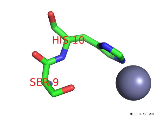
Mono view
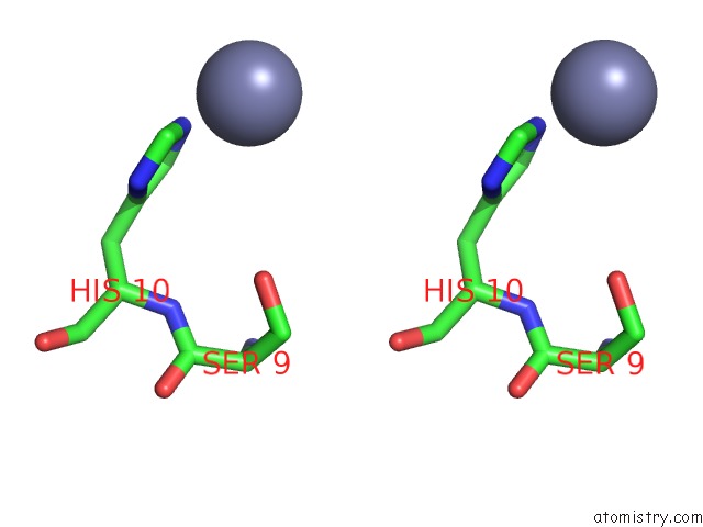
Stereo pair view
Mono view
Stereo pair view
A full contact list of Zinc with other atoms in the Zn binding
site number 1 of 3D Electron Diffraction Structure of Bovine Insulin within 5.0Å range:
|
Reference:
T.B.Blum,
D.Housset,
M.T.B.Clabbers,
E.Van Genderen,
M.Bacia-Verloop,
U.Zander,
A.A.Mccarthy,
G.Schoehn,
W.L.Ling,
J.P.Abrahams.
Statistically Correcting Dynamical Electron Scattering Improves the Refinement of Protein Nanocrystals, Including Charge Refinement of Coordinated Metals. Acta Crystallogr D Struct V. 77 75 2021BIOL.
ISSN: ISSN 2059-7983
PubMed: 33404527
DOI: 10.1107/S2059798320014540
Page generated: Tue Oct 29 15:40:54 2024
ISSN: ISSN 2059-7983
PubMed: 33404527
DOI: 10.1107/S2059798320014540
Last articles
Zn in 9J0NZn in 9J0O
Zn in 9J0P
Zn in 9FJX
Zn in 9EKB
Zn in 9C0F
Zn in 9CAH
Zn in 9CH0
Zn in 9CH3
Zn in 9CH1