Zinc »
PDB 6ogo-6oue »
6on5 »
Zinc in PDB 6on5: Crystal Structure of the Zn-Bound Domain-Swapped Dimer Q108K:T51D:A28C:L36C:F57H Mutant of Human Cellular Retinol Binding Protein II
Protein crystallography data
The structure of Crystal Structure of the Zn-Bound Domain-Swapped Dimer Q108K:T51D:A28C:L36C:F57H Mutant of Human Cellular Retinol Binding Protein II, PDB code: 6on5
was solved by
A.Ghanbarpour,
J.Geiger,
with X-Ray Crystallography technique. A brief refinement statistics is given in the table below:
| Resolution Low / High (Å) | 37.12 / 1.64 |
| Space group | P 21 21 21 |
| Cell size a, b, c (Å), α, β, γ (°) | 60.416, 61.759, 72.638, 90.00, 90.00, 90.00 |
| R / Rfree (%) | 20.4 / 25.5 |
Zinc Binding Sites:
The binding sites of Zinc atom in the Crystal Structure of the Zn-Bound Domain-Swapped Dimer Q108K:T51D:A28C:L36C:F57H Mutant of Human Cellular Retinol Binding Protein II
(pdb code 6on5). This binding sites where shown within
5.0 Angstroms radius around Zinc atom.
In total only one binding site of Zinc was determined in the Crystal Structure of the Zn-Bound Domain-Swapped Dimer Q108K:T51D:A28C:L36C:F57H Mutant of Human Cellular Retinol Binding Protein II, PDB code: 6on5:
In total only one binding site of Zinc was determined in the Crystal Structure of the Zn-Bound Domain-Swapped Dimer Q108K:T51D:A28C:L36C:F57H Mutant of Human Cellular Retinol Binding Protein II, PDB code: 6on5:
Zinc binding site 1 out of 1 in 6on5
Go back to
Zinc binding site 1 out
of 1 in the Crystal Structure of the Zn-Bound Domain-Swapped Dimer Q108K:T51D:A28C:L36C:F57H Mutant of Human Cellular Retinol Binding Protein II
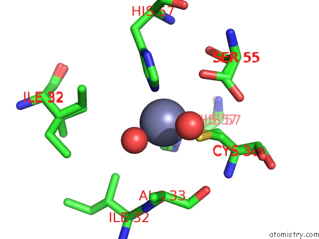
Mono view
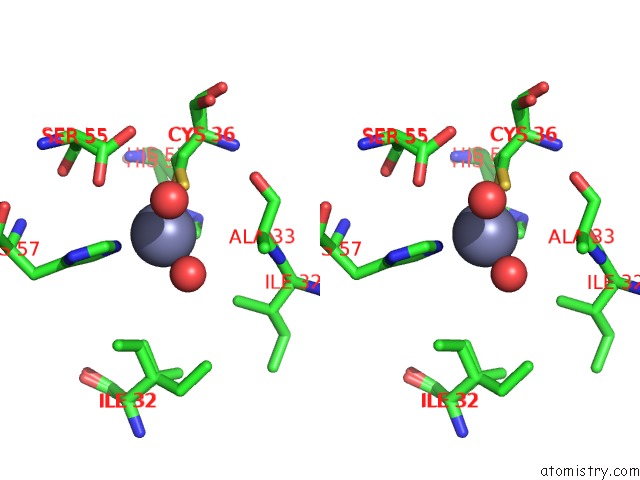
Stereo pair view
Mono view
Stereo pair view
A full contact list of Zinc with other atoms in the Zn binding
site number 1 of Crystal Structure of the Zn-Bound Domain-Swapped Dimer Q108K:T51D:A28C:L36C:F57H Mutant of Human Cellular Retinol Binding Protein II within 5.0Å range:
|
Reference:
A.Ghanbarpour,
C.Pinger,
R.Esmatpour Salmani,
Z.Assar,
E.M.Santos,
M.Nosrati,
K.Pawlowski,
D.Spence,
C.Vasileiou,
X.Jin,
B.Borhan,
J.H.Geiger.
Engineering the Hcrbpii Domain-Swapped Dimer Into A New Class of Protein Switches. J.Am.Chem.Soc. V. 141 17125 2019.
ISSN: ESSN 1520-5126
PubMed: 31557439
DOI: 10.1021/JACS.9B04664
Page generated: Tue Oct 29 04:29:16 2024
ISSN: ESSN 1520-5126
PubMed: 31557439
DOI: 10.1021/JACS.9B04664
Last articles
Zn in 9J0NZn in 9J0O
Zn in 9J0P
Zn in 9FJX
Zn in 9EKB
Zn in 9C0F
Zn in 9CAH
Zn in 9CH0
Zn in 9CH3
Zn in 9CH1