Zinc »
PDB 6go1-6h0v »
6gos »
Zinc in PDB 6gos: E. Coli Microcin Synthetase Mcbbcd Complex with Pro-MCCB17 Bound
Protein crystallography data
The structure of E. Coli Microcin Synthetase Mcbbcd Complex with Pro-MCCB17 Bound, PDB code: 6gos
was solved by
D.Ghilarov,
C.E.M.Stevenson,
D.Y.Travin,
J.Piskunova,
M.Serebryakova,
A.Maxwell,
D.M.Lawson,
K.Severinov,
with X-Ray Crystallography technique. A brief refinement statistics is given in the table below:
| Resolution Low / High (Å) | 57.39 / 2.10 |
| Space group | C 1 2 1 |
| Cell size a, b, c (Å), α, β, γ (°) | 180.960, 83.400, 86.900, 90.00, 91.45, 90.00 |
| R / Rfree (%) | 17.2 / 21.2 |
Other elements in 6gos:
The structure of E. Coli Microcin Synthetase Mcbbcd Complex with Pro-MCCB17 Bound also contains other interesting chemical elements:
| Chlorine | (Cl) | 2 atoms |
Zinc Binding Sites:
The binding sites of Zinc atom in the E. Coli Microcin Synthetase Mcbbcd Complex with Pro-MCCB17 Bound
(pdb code 6gos). This binding sites where shown within
5.0 Angstroms radius around Zinc atom.
In total 2 binding sites of Zinc where determined in the E. Coli Microcin Synthetase Mcbbcd Complex with Pro-MCCB17 Bound, PDB code: 6gos:
Jump to Zinc binding site number: 1; 2;
In total 2 binding sites of Zinc where determined in the E. Coli Microcin Synthetase Mcbbcd Complex with Pro-MCCB17 Bound, PDB code: 6gos:
Jump to Zinc binding site number: 1; 2;
Zinc binding site 1 out of 2 in 6gos
Go back to
Zinc binding site 1 out
of 2 in the E. Coli Microcin Synthetase Mcbbcd Complex with Pro-MCCB17 Bound
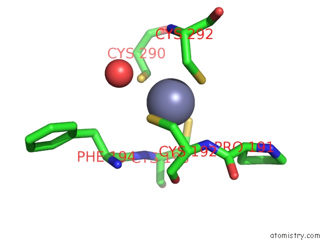
Mono view
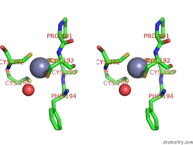
Stereo pair view
Mono view
Stereo pair view
A full contact list of Zinc with other atoms in the Zn binding
site number 1 of E. Coli Microcin Synthetase Mcbbcd Complex with Pro-MCCB17 Bound within 5.0Å range:
|
Zinc binding site 2 out of 2 in 6gos
Go back to
Zinc binding site 2 out
of 2 in the E. Coli Microcin Synthetase Mcbbcd Complex with Pro-MCCB17 Bound
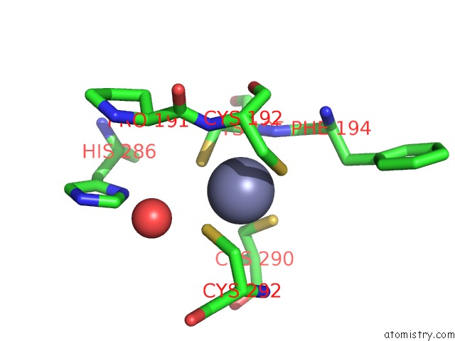
Mono view
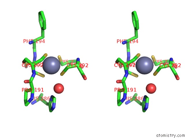
Stereo pair view
Mono view
Stereo pair view
A full contact list of Zinc with other atoms in the Zn binding
site number 2 of E. Coli Microcin Synthetase Mcbbcd Complex with Pro-MCCB17 Bound within 5.0Å range:
|
Reference:
D.Ghilarov,
C.E.M.Stevenson,
D.Y.Travin,
J.Piskunova,
M.Serebryakova,
A.Maxwell,
D.M.Lawson,
K.Severinov.
Architecture of Microcin B17 Synthetase: An Octameric Protein Complex Converting A Ribosomally Synthesized Peptide Into A Dna Gyrase Poison. Mol. Cell V. 73 749 2019.
ISSN: ISSN 1097-4164
PubMed: 30661981
DOI: 10.1016/J.MOLCEL.2018.11.032
Page generated: Mon Oct 28 21:58:02 2024
ISSN: ISSN 1097-4164
PubMed: 30661981
DOI: 10.1016/J.MOLCEL.2018.11.032
Last articles
Zn in 9J0NZn in 9J0O
Zn in 9J0P
Zn in 9FJX
Zn in 9EKB
Zn in 9C0F
Zn in 9CAH
Zn in 9CH0
Zn in 9CH3
Zn in 9CH1