Zinc »
PDB 5yy2-5zg2 »
5z98 »
Zinc in PDB 5z98: Crystal Structure of the Primate APOBEC3H Dimer Mediated By Rna Duplex
Protein crystallography data
The structure of Crystal Structure of the Primate APOBEC3H Dimer Mediated By Rna Duplex, PDB code: 5z98
was solved by
T.Matsuoka,
T.Nagae,
H.Ode,
N.Watanabe,
Y.Iwatani,
with X-Ray Crystallography technique. A brief refinement statistics is given in the table below:
| Resolution Low / High (Å) | 43.49 / 2.20 |
| Space group | P 1 21 1 |
| Cell size a, b, c (Å), α, β, γ (°) | 38.341, 86.970, 77.363, 90.00, 102.63, 90.00 |
| R / Rfree (%) | 25.1 / 32 |
Zinc Binding Sites:
The binding sites of Zinc atom in the Crystal Structure of the Primate APOBEC3H Dimer Mediated By Rna Duplex
(pdb code 5z98). This binding sites where shown within
5.0 Angstroms radius around Zinc atom.
In total 2 binding sites of Zinc where determined in the Crystal Structure of the Primate APOBEC3H Dimer Mediated By Rna Duplex, PDB code: 5z98:
Jump to Zinc binding site number: 1; 2;
In total 2 binding sites of Zinc where determined in the Crystal Structure of the Primate APOBEC3H Dimer Mediated By Rna Duplex, PDB code: 5z98:
Jump to Zinc binding site number: 1; 2;
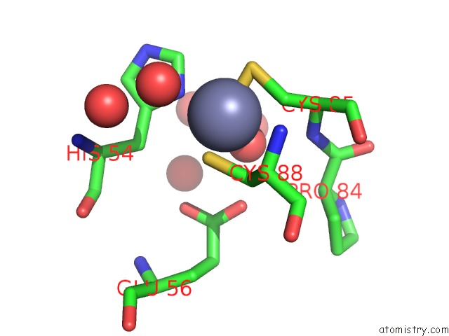
Zinc binding site 1 out of 2 in 5z98
Go back to
Zinc binding site 1 out
of 2 in the Crystal Structure of the Primate APOBEC3H Dimer Mediated By Rna Duplex
Mono view

Stereo pair view
Mono view
Stereo pair view
A full contact list of Zinc with other atoms in the Zn binding
site number 1 of Crystal Structure of the Primate APOBEC3H Dimer Mediated By Rna Duplex within 5.0Å range:
|
Zinc binding site 2 out of 2 in 5z98
Go back to
Zinc binding site 2 out
of 2 in the Crystal Structure of the Primate APOBEC3H Dimer Mediated By Rna Duplex

Mono view

Stereo pair view
Mono view
Stereo pair view
A full contact list of Zinc with other atoms in the Zn binding
site number 2 of Crystal Structure of the Primate APOBEC3H Dimer Mediated By Rna Duplex within 5.0Å range:
|
Reference:
T.Matsuoka,
T.Nagae,
H.Ode,
H.Awazu,
T.Kurosawa,
A.Hamano,
K.Matsuoka,
A.Hachiya,
M.Imahashi,
Y.Yokomaku,
N.Watanabe,
Y.Iwatani.
Structural Basis of Chimpanzee APOBEC3H Dimerization Stabilized By Double-Stranded Rna. Nucleic Acids Res. V. 46 10368 2018.
ISSN: ESSN 1362-4962
PubMed: 30060196
DOI: 10.1093/NAR/GKY676
Page generated: Mon Oct 28 16:37:18 2024
ISSN: ESSN 1362-4962
PubMed: 30060196
DOI: 10.1093/NAR/GKY676
Last articles
Zn in 9J0NZn in 9J0O
Zn in 9J0P
Zn in 9FJX
Zn in 9EKB
Zn in 9C0F
Zn in 9CAH
Zn in 9CH0
Zn in 9CH3
Zn in 9CH1