Zinc »
PDB 5fz8-5g2t »
5g11 »
Zinc in PDB 5g11: Pseudomonas Aeruginosa Hdah Bound to Pfsaha.
Protein crystallography data
The structure of Pseudomonas Aeruginosa Hdah Bound to Pfsaha., PDB code: 5g11
was solved by
A.Kraemer,
F.J.Meyer-Almes,
O.Yildiz,
with X-Ray Crystallography technique. A brief refinement statistics is given in the table below:
| Resolution Low / High (Å) | 75.96 / 2.48 |
| Space group | P 41 21 2 |
| Cell size a, b, c (Å), α, β, γ (°) | 81.790, 81.790, 204.860, 90.00, 90.00, 90.00 |
| R / Rfree (%) | 16.8 / 22.4 |
Other elements in 5g11:
The structure of Pseudomonas Aeruginosa Hdah Bound to Pfsaha. also contains other interesting chemical elements:
| Fluorine | (F) | 24 atoms |
| Potassium | (K) | 4 atoms |
Zinc Binding Sites:
The binding sites of Zinc atom in the Pseudomonas Aeruginosa Hdah Bound to Pfsaha.
(pdb code 5g11). This binding sites where shown within
5.0 Angstroms radius around Zinc atom.
In total 2 binding sites of Zinc where determined in the Pseudomonas Aeruginosa Hdah Bound to Pfsaha., PDB code: 5g11:
Jump to Zinc binding site number: 1; 2;
In total 2 binding sites of Zinc where determined in the Pseudomonas Aeruginosa Hdah Bound to Pfsaha., PDB code: 5g11:
Jump to Zinc binding site number: 1; 2;
Zinc binding site 1 out of 2 in 5g11
Go back to
Zinc binding site 1 out
of 2 in the Pseudomonas Aeruginosa Hdah Bound to Pfsaha.
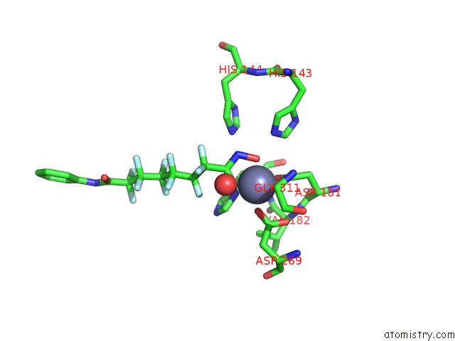
Mono view

Stereo pair view
Mono view
Stereo pair view
A full contact list of Zinc with other atoms in the Zn binding
site number 1 of Pseudomonas Aeruginosa Hdah Bound to Pfsaha. within 5.0Å range:
|
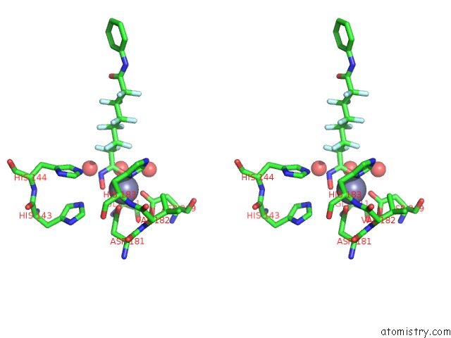
Zinc binding site 2 out of 2 in 5g11
Go back to
Zinc binding site 2 out
of 2 in the Pseudomonas Aeruginosa Hdah Bound to Pfsaha.
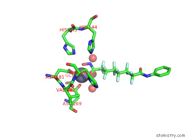
Mono view
Stereo pair view
Mono view
Stereo pair view
A full contact list of Zinc with other atoms in the Zn binding
site number 2 of Pseudomonas Aeruginosa Hdah Bound to Pfsaha. within 5.0Å range:
|
Reference:
A.Kramer,
T.Wagner,
O.Yildiz,
F.J.Meyer-Almes.
Crystal Structure of A Histone Deacetylase Homologue From Pseudomonas Aeruginosa. Biochemistry V. 55 6858 2016.
ISSN: ISSN 1520-4995
PubMed: 27951649
DOI: 10.1021/ACS.BIOCHEM.6B00613
Page generated: Sun Oct 27 16:51:24 2024
ISSN: ISSN 1520-4995
PubMed: 27951649
DOI: 10.1021/ACS.BIOCHEM.6B00613
Last articles
Zn in 9J0NZn in 9J0O
Zn in 9J0P
Zn in 9FJX
Zn in 9EKB
Zn in 9C0F
Zn in 9CAH
Zn in 9CH0
Zn in 9CH3
Zn in 9CH1