Zinc »
PDB 5cdg-5cup »
5cpa »
Zinc in PDB 5cpa: Refined Crystal Structure of Carboxypeptidase A at 1.54 Angstroms Resolution.
Enzymatic activity of Refined Crystal Structure of Carboxypeptidase A at 1.54 Angstroms Resolution.
All present enzymatic activity of Refined Crystal Structure of Carboxypeptidase A at 1.54 Angstroms Resolution.:
3.4.17.1;
3.4.17.1;
Protein crystallography data
The structure of Refined Crystal Structure of Carboxypeptidase A at 1.54 Angstroms Resolution., PDB code: 5cpa
was solved by
W.N.Lipscomb,
with X-Ray Crystallography technique. A brief refinement statistics is given in the table below:
| Resolution Low / High (Å) | N/A / 1.54 |
| Space group | P 1 21 1 |
| Cell size a, b, c (Å), α, β, γ (°) | 51.600, 60.270, 47.250, 90.00, 97.27, 90.00 |
| R / Rfree (%) | n/a / n/a |
Zinc Binding Sites:
The binding sites of Zinc atom in the Refined Crystal Structure of Carboxypeptidase A at 1.54 Angstroms Resolution.
(pdb code 5cpa). This binding sites where shown within
5.0 Angstroms radius around Zinc atom.
In total only one binding site of Zinc was determined in the Refined Crystal Structure of Carboxypeptidase A at 1.54 Angstroms Resolution., PDB code: 5cpa:
In total only one binding site of Zinc was determined in the Refined Crystal Structure of Carboxypeptidase A at 1.54 Angstroms Resolution., PDB code: 5cpa:
Zinc binding site 1 out of 1 in 5cpa
Go back to
Zinc binding site 1 out
of 1 in the Refined Crystal Structure of Carboxypeptidase A at 1.54 Angstroms Resolution.
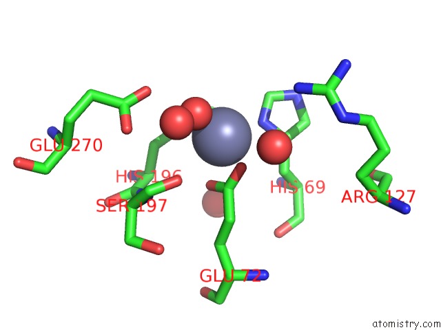
Mono view
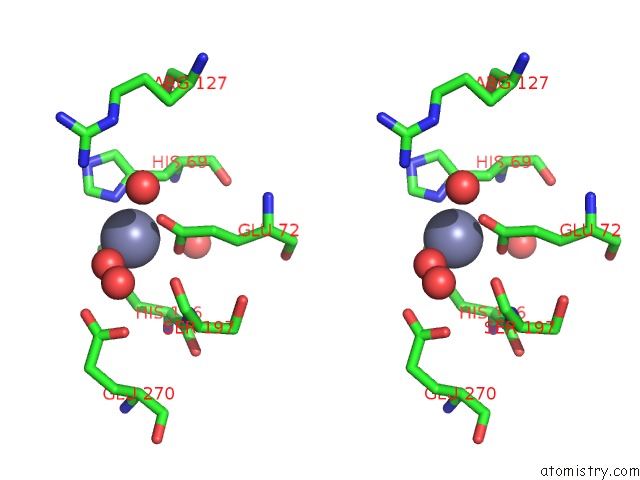
Stereo pair view
Mono view
Stereo pair view
A full contact list of Zinc with other atoms in the Zn binding
site number 1 of Refined Crystal Structure of Carboxypeptidase A at 1.54 Angstroms Resolution. within 5.0Å range:
|
Reference:
D.C.Rees,
M.Lewis,
W.N.Lipscomb.
Refined Crystal Structure of Carboxypeptidase A at 1.54 A Resolution. J.Mol.Biol. V. 168 367 1983.
ISSN: ISSN 0022-2836
PubMed: 6887246
DOI: 10.1016/S0022-2836(83)80024-2
Page generated: Sun Oct 27 14:26:07 2024
ISSN: ISSN 0022-2836
PubMed: 6887246
DOI: 10.1016/S0022-2836(83)80024-2
Last articles
Zn in 9JPJZn in 9JP7
Zn in 9JPK
Zn in 9JPL
Zn in 9GN6
Zn in 9GN7
Zn in 9GKU
Zn in 9GKW
Zn in 9GKX
Zn in 9GL0