Zinc »
PDB 3dgn-3dsw »
3dra »
Zinc in PDB 3dra: Candida Albicans Protein Geranylgeranyltransferase-I Complexed with Ggpp
Enzymatic activity of Candida Albicans Protein Geranylgeranyltransferase-I Complexed with Ggpp
All present enzymatic activity of Candida Albicans Protein Geranylgeranyltransferase-I Complexed with Ggpp:
2.5.1.58; 2.5.1.59;
2.5.1.58; 2.5.1.59;
Protein crystallography data
The structure of Candida Albicans Protein Geranylgeranyltransferase-I Complexed with Ggpp, PDB code: 3dra
was solved by
M.A.Hast,
L.S.Beese,
with X-Ray Crystallography technique. A brief refinement statistics is given in the table below:
| Resolution Low / High (Å) | 37.64 / 1.80 |
| Space group | C 1 2 1 |
| Cell size a, b, c (Å), α, β, γ (°) | 132.304, 66.049, 82.821, 90.00, 100.02, 90.00 |
| R / Rfree (%) | 19.2 / 23.1 |
Zinc Binding Sites:
The binding sites of Zinc atom in the Candida Albicans Protein Geranylgeranyltransferase-I Complexed with Ggpp
(pdb code 3dra). This binding sites where shown within
5.0 Angstroms radius around Zinc atom.
In total only one binding site of Zinc was determined in the Candida Albicans Protein Geranylgeranyltransferase-I Complexed with Ggpp, PDB code: 3dra:
In total only one binding site of Zinc was determined in the Candida Albicans Protein Geranylgeranyltransferase-I Complexed with Ggpp, PDB code: 3dra:
Zinc binding site 1 out of 1 in 3dra
Go back to
Zinc binding site 1 out
of 1 in the Candida Albicans Protein Geranylgeranyltransferase-I Complexed with Ggpp
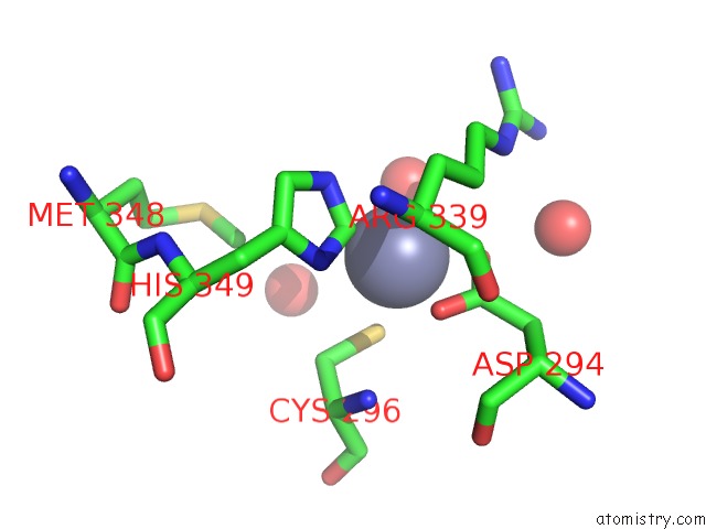
Mono view
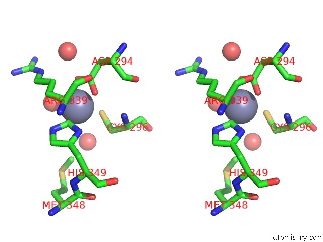
Stereo pair view
Mono view
Stereo pair view
A full contact list of Zinc with other atoms in the Zn binding
site number 1 of Candida Albicans Protein Geranylgeranyltransferase-I Complexed with Ggpp within 5.0Å range:
|
Reference:
M.A.Hast,
L.S.Beese.
Structure of Protein Geranylgeranyltransferase-I From the Human Pathogen Candida Albicans Complexed with A Lipid Substrate. J.Biol.Chem. V. 283 31933 2008.
ISSN: ISSN 0021-9258
PubMed: 18713740
DOI: 10.1074/JBC.M805330200
Page generated: Thu Oct 24 12:19:06 2024
ISSN: ISSN 0021-9258
PubMed: 18713740
DOI: 10.1074/JBC.M805330200
Last articles
Zn in 9J0NZn in 9J0O
Zn in 9J0P
Zn in 9FJX
Zn in 9EKB
Zn in 9C0F
Zn in 9CAH
Zn in 9CH0
Zn in 9CH3
Zn in 9CH1