Zinc »
PDB 3dgn-3dsw »
3dln »
Zinc in PDB 3dln: Crystal Structure of the Binding Domain of the Ampa Subunit GLUR3 Bound to Glutamate
Protein crystallography data
The structure of Crystal Structure of the Binding Domain of the Ampa Subunit GLUR3 Bound to Glutamate, PDB code: 3dln
was solved by
A.H.Ahmed,
Q.Wang,
H.Sondermann,
R.E.Oswald,
with X-Ray Crystallography technique. A brief refinement statistics is given in the table below:
| Resolution Low / High (Å) | 50.00 / 1.91 |
| Space group | P 2 2 21 |
| Cell size a, b, c (Å), α, β, γ (°) | 47.585, 47.402, 137.940, 90.00, 90.00, 90.00 |
| R / Rfree (%) | 19.7 / 23.5 |
Zinc Binding Sites:
The binding sites of Zinc atom in the Crystal Structure of the Binding Domain of the Ampa Subunit GLUR3 Bound to Glutamate
(pdb code 3dln). This binding sites where shown within
5.0 Angstroms radius around Zinc atom.
In total only one binding site of Zinc was determined in the Crystal Structure of the Binding Domain of the Ampa Subunit GLUR3 Bound to Glutamate, PDB code: 3dln:
In total only one binding site of Zinc was determined in the Crystal Structure of the Binding Domain of the Ampa Subunit GLUR3 Bound to Glutamate, PDB code: 3dln:
Zinc binding site 1 out of 1 in 3dln
Go back to
Zinc binding site 1 out
of 1 in the Crystal Structure of the Binding Domain of the Ampa Subunit GLUR3 Bound to Glutamate
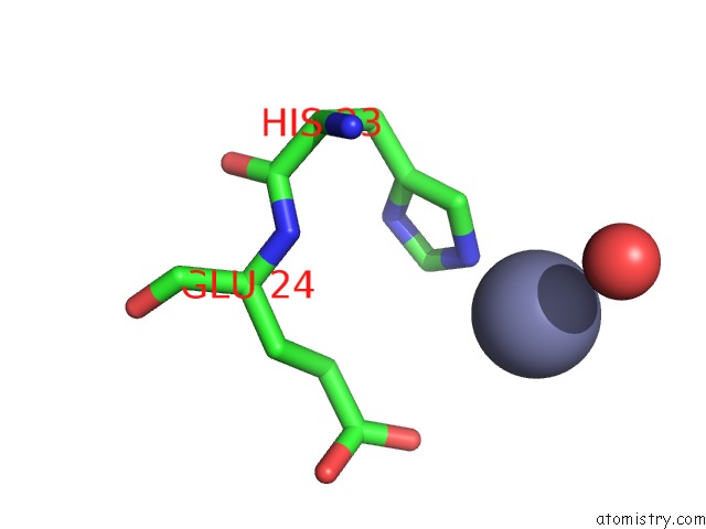
Mono view
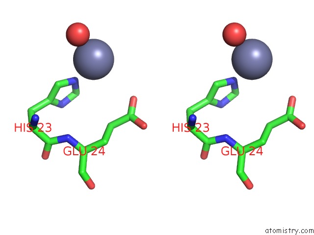
Stereo pair view
Mono view
Stereo pair view
A full contact list of Zinc with other atoms in the Zn binding
site number 1 of Crystal Structure of the Binding Domain of the Ampa Subunit GLUR3 Bound to Glutamate within 5.0Å range:
|
Reference:
A.H.Ahmed,
Q.Wang,
H.Sondermann,
R.E.Oswald.
Structure of the S1S2 Glutamate Binding Domain of GLUR3. Proteins V. 75 628 2008.
ISSN: ISSN 0887-3585
PubMed: 19003990
DOI: 10.1002/PROT.22274
Page generated: Thu Oct 24 12:15:33 2024
ISSN: ISSN 0887-3585
PubMed: 19003990
DOI: 10.1002/PROT.22274
Last articles
Zn in 9J0NZn in 9J0O
Zn in 9J0P
Zn in 9FJX
Zn in 9EKB
Zn in 9C0F
Zn in 9CAH
Zn in 9CH0
Zn in 9CH3
Zn in 9CH1