Zinc »
PDB 3dgn-3dsw »
3dhb »
Zinc in PDB 3dhb: 1.4 Angstrom Structure of N-Acyl Homoserine Lactone Hydrolase with the Product N-Hexanoyl-L-Homoserine Bound at the Catalytic Metal Center
Protein crystallography data
The structure of 1.4 Angstrom Structure of N-Acyl Homoserine Lactone Hydrolase with the Product N-Hexanoyl-L-Homoserine Bound at the Catalytic Metal Center, PDB code: 3dhb
was solved by
D.Liu,
J.Momb,
P.W.Thomas,
A.Moulin,
G.A.Petsko,
W.Fast,
D.Ringe,
with X-Ray Crystallography technique. A brief refinement statistics is given in the table below:
| Resolution Low / High (Å) | 18.79 / 1.40 |
| Space group | P 21 21 21 |
| Cell size a, b, c (Å), α, β, γ (°) | 55.438, 55.583, 79.866, 90.00, 90.00, 90.00 |
| R / Rfree (%) | 14.2 / 18.4 |
Zinc Binding Sites:
The binding sites of Zinc atom in the 1.4 Angstrom Structure of N-Acyl Homoserine Lactone Hydrolase with the Product N-Hexanoyl-L-Homoserine Bound at the Catalytic Metal Center
(pdb code 3dhb). This binding sites where shown within
5.0 Angstroms radius around Zinc atom.
In total 2 binding sites of Zinc where determined in the 1.4 Angstrom Structure of N-Acyl Homoserine Lactone Hydrolase with the Product N-Hexanoyl-L-Homoserine Bound at the Catalytic Metal Center, PDB code: 3dhb:
Jump to Zinc binding site number: 1; 2;
In total 2 binding sites of Zinc where determined in the 1.4 Angstrom Structure of N-Acyl Homoserine Lactone Hydrolase with the Product N-Hexanoyl-L-Homoserine Bound at the Catalytic Metal Center, PDB code: 3dhb:
Jump to Zinc binding site number: 1; 2;
Zinc binding site 1 out of 2 in 3dhb
Go back to
Zinc binding site 1 out
of 2 in the 1.4 Angstrom Structure of N-Acyl Homoserine Lactone Hydrolase with the Product N-Hexanoyl-L-Homoserine Bound at the Catalytic Metal Center
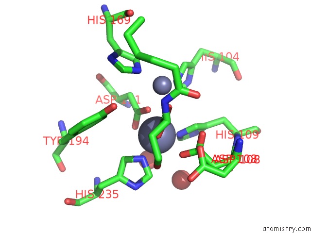
Mono view
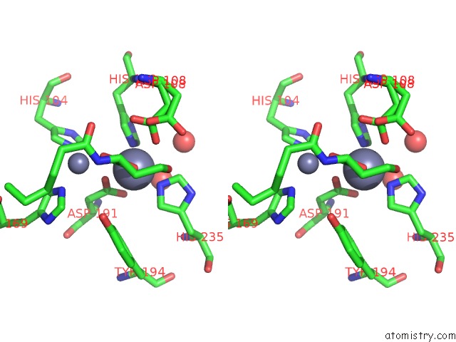
Stereo pair view
Mono view
Stereo pair view
A full contact list of Zinc with other atoms in the Zn binding
site number 1 of 1.4 Angstrom Structure of N-Acyl Homoserine Lactone Hydrolase with the Product N-Hexanoyl-L-Homoserine Bound at the Catalytic Metal Center within 5.0Å range:
|
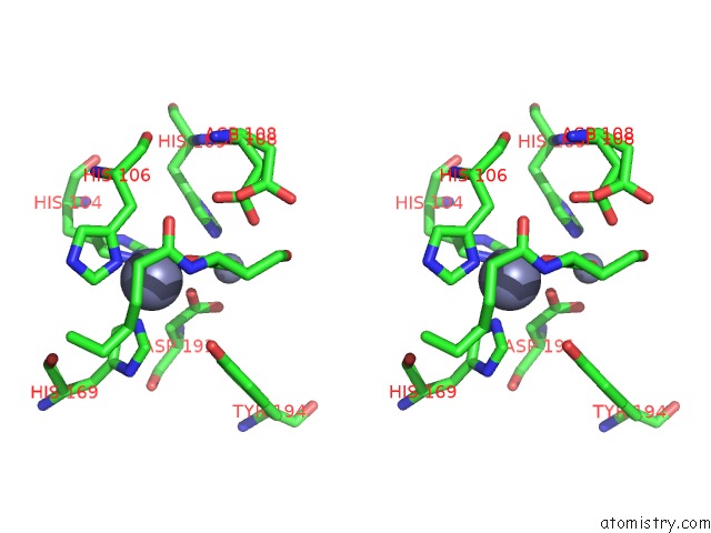
Zinc binding site 2 out of 2 in 3dhb
Go back to
Zinc binding site 2 out
of 2 in the 1.4 Angstrom Structure of N-Acyl Homoserine Lactone Hydrolase with the Product N-Hexanoyl-L-Homoserine Bound at the Catalytic Metal Center
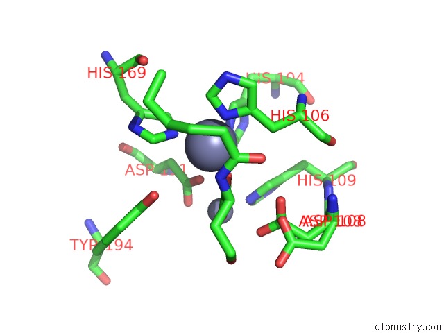
Mono view
Stereo pair view
Mono view
Stereo pair view
A full contact list of Zinc with other atoms in the Zn binding
site number 2 of 1.4 Angstrom Structure of N-Acyl Homoserine Lactone Hydrolase with the Product N-Hexanoyl-L-Homoserine Bound at the Catalytic Metal Center within 5.0Å range:
|
Reference:
D.Liu,
J.Momb,
P.W.Thomas,
A.Moulin,
G.A.Petsko,
W.Fast,
D.Ringe.
Mechanism of the Quorum-Quenching Lactonase (Aiia) From Bacillus Thuringiensis. 1. Product-Bound Structures. Biochemistry V. 47 7706 2008.
ISSN: ISSN 0006-2960
PubMed: 18627129
DOI: 10.1021/BI800368Y
Page generated: Thu Oct 24 12:13:24 2024
ISSN: ISSN 0006-2960
PubMed: 18627129
DOI: 10.1021/BI800368Y
Last articles
Zn in 9J0NZn in 9J0O
Zn in 9J0P
Zn in 9FJX
Zn in 9EKB
Zn in 9C0F
Zn in 9CAH
Zn in 9CH0
Zn in 9CH3
Zn in 9CH1