Zinc »
PDB 3dgn-3dsw »
3dgv »
Zinc in PDB 3dgv: Crystal Structure of Thrombin Activatable Fibrinolysis Inhibitor (Tafi)
Enzymatic activity of Crystal Structure of Thrombin Activatable Fibrinolysis Inhibitor (Tafi)
All present enzymatic activity of Crystal Structure of Thrombin Activatable Fibrinolysis Inhibitor (Tafi):
3.4.17.20;
3.4.17.20;
Protein crystallography data
The structure of Crystal Structure of Thrombin Activatable Fibrinolysis Inhibitor (Tafi), PDB code: 3dgv
was solved by
K.Anand,
I.Pallares,
Z.Valnickova,
T.Christensen,
H.Schreuder,
J.Enghild,
with X-Ray Crystallography technique. A brief refinement statistics is given in the table below:
| Resolution Low / High (Å) | 19.86 / 2.50 |
| Space group | P 41 21 2 |
| Cell size a, b, c (Å), α, β, γ (°) | 146.470, 146.470, 231.690, 90.00, 90.00, 90.00 |
| R / Rfree (%) | 20.5 / 24.3 |
Zinc Binding Sites:
The binding sites of Zinc atom in the Crystal Structure of Thrombin Activatable Fibrinolysis Inhibitor (Tafi)
(pdb code 3dgv). This binding sites where shown within
5.0 Angstroms radius around Zinc atom.
In total 3 binding sites of Zinc where determined in the Crystal Structure of Thrombin Activatable Fibrinolysis Inhibitor (Tafi), PDB code: 3dgv:
Jump to Zinc binding site number: 1; 2; 3;
In total 3 binding sites of Zinc where determined in the Crystal Structure of Thrombin Activatable Fibrinolysis Inhibitor (Tafi), PDB code: 3dgv:
Jump to Zinc binding site number: 1; 2; 3;
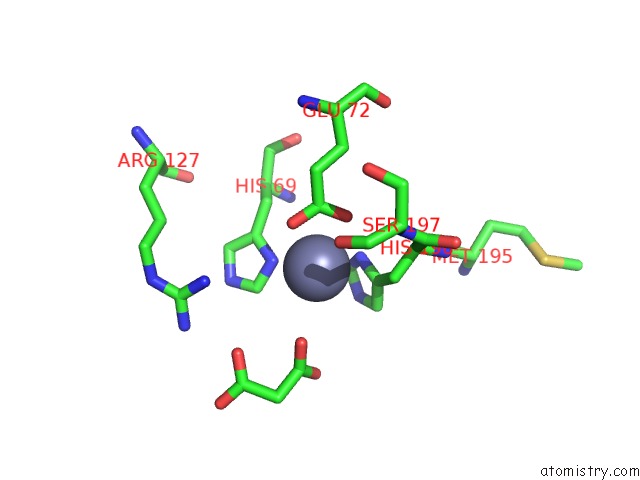
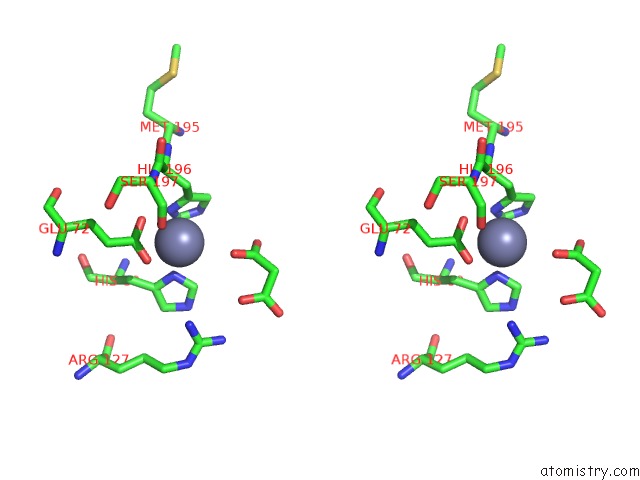
Zinc binding site 1 out of 3 in 3dgv
Go back to
Zinc binding site 1 out
of 3 in the Crystal Structure of Thrombin Activatable Fibrinolysis Inhibitor (Tafi)
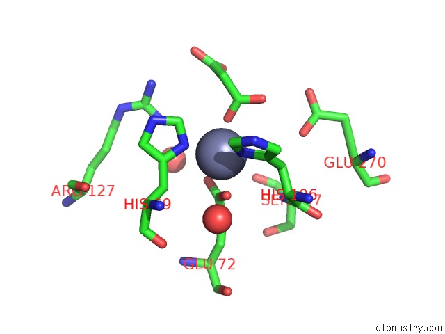
Mono view
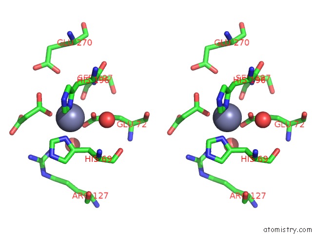
Stereo pair view
Mono view
Stereo pair view
A full contact list of Zinc with other atoms in the Zn binding
site number 1 of Crystal Structure of Thrombin Activatable Fibrinolysis Inhibitor (Tafi) within 5.0Å range:
|
Zinc binding site 2 out of 3 in 3dgv
Go back to
Zinc binding site 2 out
of 3 in the Crystal Structure of Thrombin Activatable Fibrinolysis Inhibitor (Tafi)
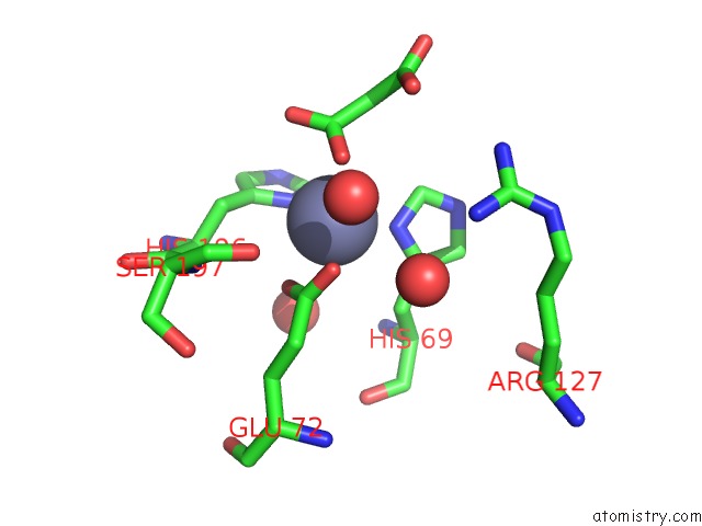
Mono view
Stereo pair view
Mono view
Stereo pair view
A full contact list of Zinc with other atoms in the Zn binding
site number 2 of Crystal Structure of Thrombin Activatable Fibrinolysis Inhibitor (Tafi) within 5.0Å range:
|
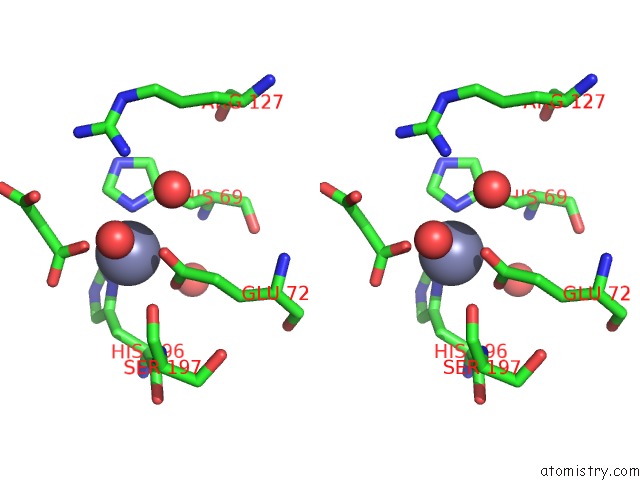
Zinc binding site 3 out of 3 in 3dgv
Go back to
Zinc binding site 3 out
of 3 in the Crystal Structure of Thrombin Activatable Fibrinolysis Inhibitor (Tafi)
Mono view
Stereo pair view
Mono view
Stereo pair view
A full contact list of Zinc with other atoms in the Zn binding
site number 3 of Crystal Structure of Thrombin Activatable Fibrinolysis Inhibitor (Tafi) within 5.0Å range:
|
Reference:
K.Anand,
I.Pallares,
Z.Valnickova,
T.Christensen,
J.Vendrell,
K.U.Wendt,
H.A.Schreuder,
J.J.Enghild,
F.X.Avil S.
The Crystal Structure of Tafi Provides the Structural Basis For the Intrinsic Activity of the Proenzyme and Short Half-Life of Tafia To Be Published.
Page generated: Thu Oct 24 12:13:23 2024
Last articles
Zn in 9J0NZn in 9J0O
Zn in 9J0P
Zn in 9FJX
Zn in 9EKB
Zn in 9C0F
Zn in 9CAH
Zn in 9CH0
Zn in 9CH3
Zn in 9CH1