Zinc »
PDB 2q42-2qmu »
2q8j »
Zinc in PDB 2q8j: Crystal Structure of the Complex of C-Lobe of Bovine Lactoferrin with Mannitol and Mannose at 2.7 A Resolution
Protein crystallography data
The structure of Crystal Structure of the Complex of C-Lobe of Bovine Lactoferrin with Mannitol and Mannose at 2.7 A Resolution, PDB code: 2q8j
was solved by
R.Mir,
R.Jain,
M.Sinha,
N.Singh,
S.Sharma,
P.Kaur,
A.Bhushan,
T.P.Singh,
with X-Ray Crystallography technique. A brief refinement statistics is given in the table below:
| Resolution Low / High (Å) | 20.00 / 2.71 |
| Space group | P 1 21 1 |
| Cell size a, b, c (Å), α, β, γ (°) | 63.400, 50.300, 65.900, 90.00, 107.80, 90.00 |
| R / Rfree (%) | 19.5 / 24 |
Other elements in 2q8j:
The structure of Crystal Structure of the Complex of C-Lobe of Bovine Lactoferrin with Mannitol and Mannose at 2.7 A Resolution also contains other interesting chemical elements:
| Iron | (Fe) | 1 atom |
Zinc Binding Sites:
The binding sites of Zinc atom in the Crystal Structure of the Complex of C-Lobe of Bovine Lactoferrin with Mannitol and Mannose at 2.7 A Resolution
(pdb code 2q8j). This binding sites where shown within
5.0 Angstroms radius around Zinc atom.
In total 2 binding sites of Zinc where determined in the Crystal Structure of the Complex of C-Lobe of Bovine Lactoferrin with Mannitol and Mannose at 2.7 A Resolution, PDB code: 2q8j:
Jump to Zinc binding site number: 1; 2;
In total 2 binding sites of Zinc where determined in the Crystal Structure of the Complex of C-Lobe of Bovine Lactoferrin with Mannitol and Mannose at 2.7 A Resolution, PDB code: 2q8j:
Jump to Zinc binding site number: 1; 2;
Zinc binding site 1 out of 2 in 2q8j
Go back to
Zinc binding site 1 out
of 2 in the Crystal Structure of the Complex of C-Lobe of Bovine Lactoferrin with Mannitol and Mannose at 2.7 A Resolution
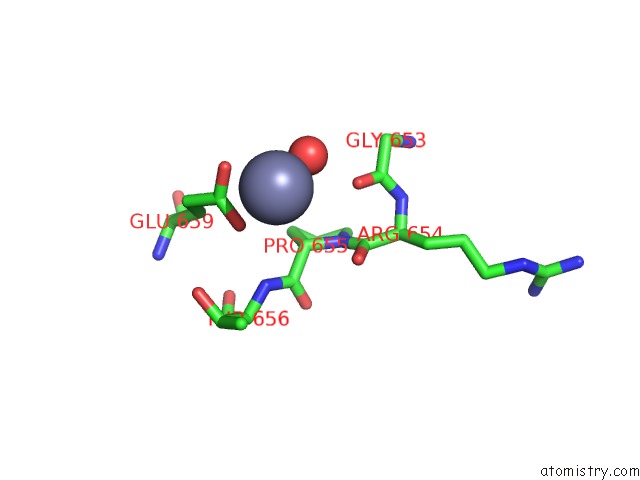
Mono view
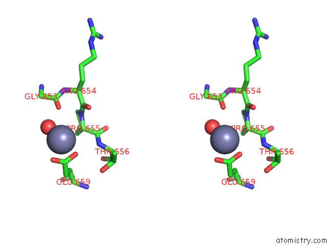
Stereo pair view
Mono view
Stereo pair view
A full contact list of Zinc with other atoms in the Zn binding
site number 1 of Crystal Structure of the Complex of C-Lobe of Bovine Lactoferrin with Mannitol and Mannose at 2.7 A Resolution within 5.0Å range:
|
Zinc binding site 2 out of 2 in 2q8j
Go back to
Zinc binding site 2 out
of 2 in the Crystal Structure of the Complex of C-Lobe of Bovine Lactoferrin with Mannitol and Mannose at 2.7 A Resolution
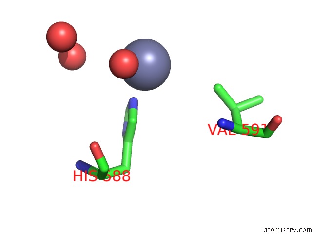
Mono view
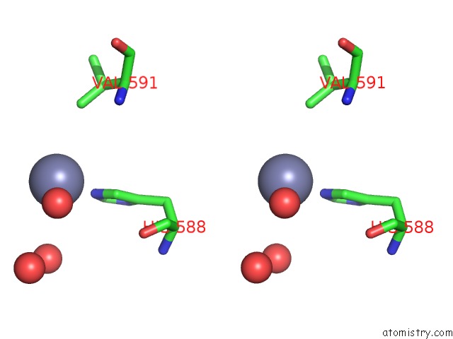
Stereo pair view
Mono view
Stereo pair view
A full contact list of Zinc with other atoms in the Zn binding
site number 2 of Crystal Structure of the Complex of C-Lobe of Bovine Lactoferrin with Mannitol and Mannose at 2.7 A Resolution within 5.0Å range:
|
Reference:
R.Mir,
R.Jain,
M.Sinha,
N.Singh,
S.Sharma,
P.Kaur,
A.Bhushan,
T.P.Singh.
Crystal Structure of the Complex of C-Lobe of Bovine Lactoferrin with Mannitol and Mannose at 2.7 A Resolution To Be Published.
Page generated: Wed Aug 20 05:21:10 2025
Last articles
Zn in 3H3KZn in 3H2Q
Zn in 3H3E
Zn in 3H2P
Zn in 3H2W
Zn in 3H1Y
Zn in 3H1W
Zn in 3H1K
Zn in 3H0G
Zn in 3H1M