Zinc »
PDB 3gtv-3h68 »
3h2q »
Zinc in PDB 3h2q: Human SOD1 H80R Variant, P21 Crystal Form
Enzymatic activity of Human SOD1 H80R Variant, P21 Crystal Form
All present enzymatic activity of Human SOD1 H80R Variant, P21 Crystal Form:
1.15.1.1;
1.15.1.1;
Protein crystallography data
The structure of Human SOD1 H80R Variant, P21 Crystal Form, PDB code: 3h2q
was solved by
S.V.Seetharaman,
D.D.Winkler,
A.B.Taylor,
X.Cao,
L.J.Whitson,
P.A.Doucette,
J.S.Valentine,
M.C.Carroll,
V.C.Culotta,
P.J.Hart,
with X-Ray Crystallography technique. A brief refinement statistics is given in the table below:
| Resolution Low / High (Å) | 25.34 / 1.85 |
| Space group | P 1 21 1 |
| Cell size a, b, c (Å), α, β, γ (°) | 35.240, 136.801, 56.364, 90.00, 104.58, 90.00 |
| R / Rfree (%) | 17.9 / 22.2 |
Zinc Binding Sites:
The binding sites of Zinc atom in the Human SOD1 H80R Variant, P21 Crystal Form
(pdb code 3h2q). This binding sites where shown within
5.0 Angstroms radius around Zinc atom.
In total 4 binding sites of Zinc where determined in the Human SOD1 H80R Variant, P21 Crystal Form, PDB code: 3h2q:
Jump to Zinc binding site number: 1; 2; 3; 4;
In total 4 binding sites of Zinc where determined in the Human SOD1 H80R Variant, P21 Crystal Form, PDB code: 3h2q:
Jump to Zinc binding site number: 1; 2; 3; 4;
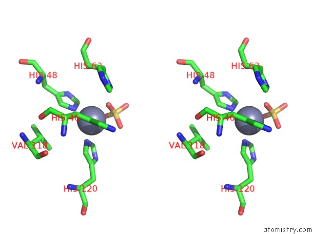
Zinc binding site 1 out of 4 in 3h2q
Go back to
Zinc binding site 1 out
of 4 in the Human SOD1 H80R Variant, P21 Crystal Form
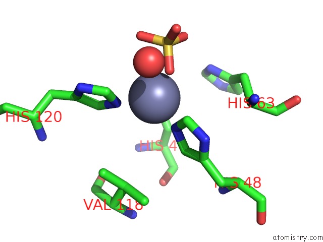
Mono view

Stereo pair view
Mono view
Stereo pair view
A full contact list of Zinc with other atoms in the Zn binding
site number 1 of Human SOD1 H80R Variant, P21 Crystal Form within 5.0Å range:
|
Zinc binding site 2 out of 4 in 3h2q
Go back to
Zinc binding site 2 out
of 4 in the Human SOD1 H80R Variant, P21 Crystal Form

Mono view

Stereo pair view
Mono view
Stereo pair view
A full contact list of Zinc with other atoms in the Zn binding
site number 2 of Human SOD1 H80R Variant, P21 Crystal Form within 5.0Å range:
|
Zinc binding site 3 out of 4 in 3h2q
Go back to
Zinc binding site 3 out
of 4 in the Human SOD1 H80R Variant, P21 Crystal Form

Mono view

Stereo pair view
Mono view
Stereo pair view
A full contact list of Zinc with other atoms in the Zn binding
site number 3 of Human SOD1 H80R Variant, P21 Crystal Form within 5.0Å range:
|
Zinc binding site 4 out of 4 in 3h2q
Go back to
Zinc binding site 4 out
of 4 in the Human SOD1 H80R Variant, P21 Crystal Form

Mono view
Stereo pair view
Mono view
Stereo pair view
A full contact list of Zinc with other atoms in the Zn binding
site number 4 of Human SOD1 H80R Variant, P21 Crystal Form within 5.0Å range:
|
Reference:
S.V.Seetharaman,
D.D.Winkler,
A.B.Taylor,
X.Cao,
L.J.Whitson,
P.A.Doucette,
J.S.Valentine,
M.C.Carroll,
V.C.Culotta,
P.J.Hart.
Structures of Pathogenic SOD1 Mutants H80R and D124V: Disrupted Zinc-Binding and Compromised Post-Translational Modification By the Copper Chaperone Ccs To Be Published.
Page generated: Thu Oct 24 14:11:31 2024
Last articles
Mg in 5OCHMg in 5OCO
Mg in 5OCM
Mg in 5OCG
Mg in 5OC0
Mg in 5OBY
Mg in 5OAT
Mg in 5OBW
Mg in 5OBU
Mg in 5OBJ