Zinc »
PDB 1z5r-1zfo »
1z6r »
Zinc in PDB 1z6r: Crystal Structure of Mlc From Escherichia Coli
Protein crystallography data
The structure of Crystal Structure of Mlc From Escherichia Coli, PDB code: 1z6r
was solved by
A.Schiefner,
K.Gerber,
S.Seitz,
W.Welte,
K.Diederichs,
W.Boos,
with X-Ray Crystallography technique. A brief refinement statistics is given in the table below:
| Resolution Low / High (Å) | 19.94 / 2.70 |
| Space group | C 1 2 1 |
| Cell size a, b, c (Å), α, β, γ (°) | 235.950, 74.710, 154.950, 90.00, 129.15, 90.00 |
| R / Rfree (%) | 20.3 / 26.3 |
Zinc Binding Sites:
The binding sites of Zinc atom in the Crystal Structure of Mlc From Escherichia Coli
(pdb code 1z6r). This binding sites where shown within
5.0 Angstroms radius around Zinc atom.
In total 4 binding sites of Zinc where determined in the Crystal Structure of Mlc From Escherichia Coli, PDB code: 1z6r:
Jump to Zinc binding site number: 1; 2; 3; 4;
In total 4 binding sites of Zinc where determined in the Crystal Structure of Mlc From Escherichia Coli, PDB code: 1z6r:
Jump to Zinc binding site number: 1; 2; 3; 4;
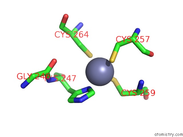
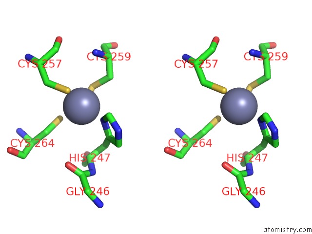
Zinc binding site 1 out of 4 in 1z6r
Go back to
Zinc binding site 1 out
of 4 in the Crystal Structure of Mlc From Escherichia Coli
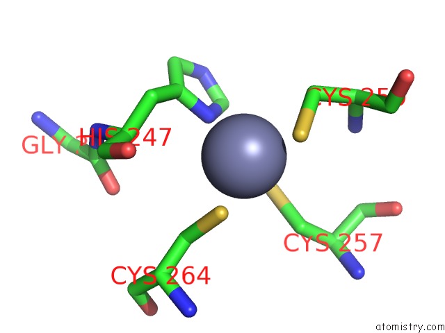
Mono view
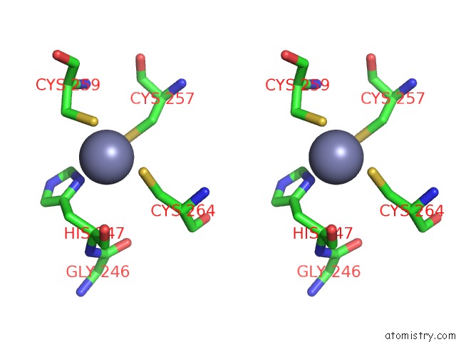
Stereo pair view
Mono view
Stereo pair view
A full contact list of Zinc with other atoms in the Zn binding
site number 1 of Crystal Structure of Mlc From Escherichia Coli within 5.0Å range:
|
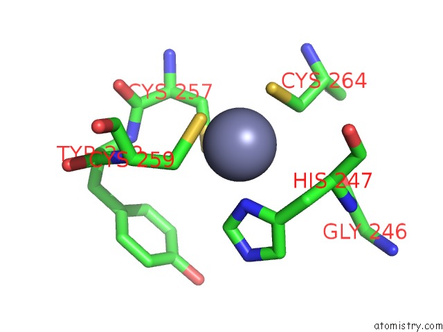
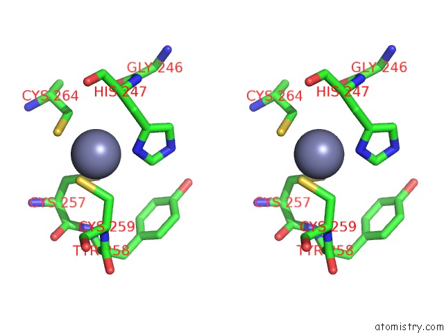
Zinc binding site 2 out of 4 in 1z6r
Go back to
Zinc binding site 2 out
of 4 in the Crystal Structure of Mlc From Escherichia Coli
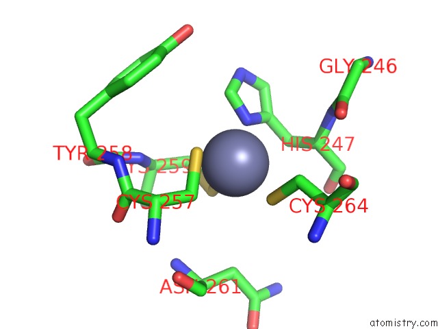
Mono view
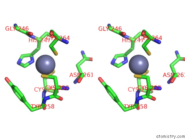
Stereo pair view
Mono view
Stereo pair view
A full contact list of Zinc with other atoms in the Zn binding
site number 2 of Crystal Structure of Mlc From Escherichia Coli within 5.0Å range:
|
Zinc binding site 3 out of 4 in 1z6r
Go back to
Zinc binding site 3 out
of 4 in the Crystal Structure of Mlc From Escherichia Coli
Mono view
Stereo pair view
Mono view
Stereo pair view
A full contact list of Zinc with other atoms in the Zn binding
site number 3 of Crystal Structure of Mlc From Escherichia Coli within 5.0Å range:
|
Zinc binding site 4 out of 4 in 1z6r
Go back to
Zinc binding site 4 out
of 4 in the Crystal Structure of Mlc From Escherichia Coli
Mono view
Stereo pair view
Mono view
Stereo pair view
A full contact list of Zinc with other atoms in the Zn binding
site number 4 of Crystal Structure of Mlc From Escherichia Coli within 5.0Å range:
|
Reference:
A.Schiefner,
K.Gerber,
S.Seitz,
W.Welte,
K.Diederichs,
W.Boos.
The Crystal Structure of Mlc, A Global Regulator of Sugar Metabolism in Escherichia Coli J.Biol.Chem. V. 280 29073 2005.
ISSN: ISSN 0021-9258
PubMed: 15929984
DOI: 10.1074/JBC.M504215200
Page generated: Wed Aug 20 00:47:51 2025
ISSN: ISSN 0021-9258
PubMed: 15929984
DOI: 10.1074/JBC.M504215200
Last articles
Zn in 2MUMZn in 2MUR
Zn in 2MUQ
Zn in 2MS0
Zn in 2MS1
Zn in 2MS3
Zn in 2MQ1
Zn in 2MRF
Zn in 2MQV
Zn in 2MRE