Zinc »
PDB 1x8h-1xm8 »
1xa8 »
Zinc in PDB 1xa8: Crystal Structure Analysis of Glutathione-Dependent Formaldehyde- Activating Enzyme (Gfa)
Protein crystallography data
The structure of Crystal Structure Analysis of Glutathione-Dependent Formaldehyde- Activating Enzyme (Gfa), PDB code: 1xa8
was solved by
A.M.Neculai,
D.Neculai,
C.Griesinger,
J.A.Vorholt,
S.Becker,
with X-Ray Crystallography technique. A brief refinement statistics is given in the table below:
| Resolution Low / High (Å) | 75.16 / 2.40 |
| Space group | P 1 21 1 |
| Cell size a, b, c (Å), α, β, γ (°) | 53.910, 120.620, 97.140, 90.00, 97.68, 90.00 |
| R / Rfree (%) | 19.5 / 24.8 |
Zinc Binding Sites:
The binding sites of Zinc atom in the Crystal Structure Analysis of Glutathione-Dependent Formaldehyde- Activating Enzyme (Gfa)
(pdb code 1xa8). This binding sites where shown within
5.0 Angstroms radius around Zinc atom.
In total 4 binding sites of Zinc where determined in the Crystal Structure Analysis of Glutathione-Dependent Formaldehyde- Activating Enzyme (Gfa), PDB code: 1xa8:
Jump to Zinc binding site number: 1; 2; 3; 4;
In total 4 binding sites of Zinc where determined in the Crystal Structure Analysis of Glutathione-Dependent Formaldehyde- Activating Enzyme (Gfa), PDB code: 1xa8:
Jump to Zinc binding site number: 1; 2; 3; 4;
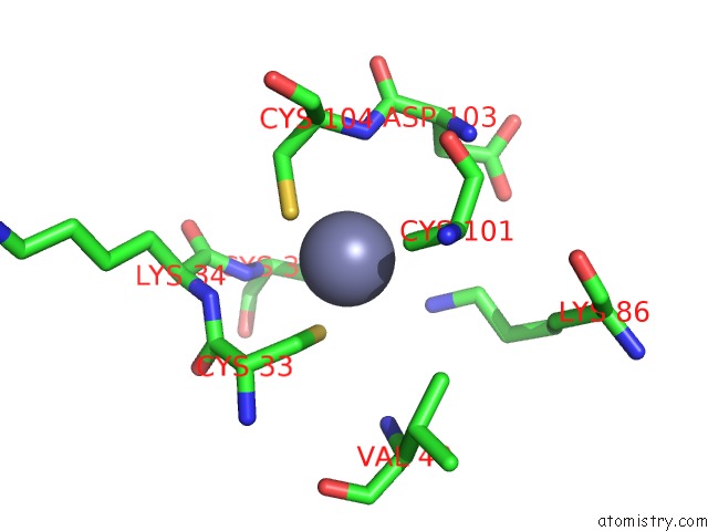
Zinc binding site 1 out of 4 in 1xa8
Go back to
Zinc binding site 1 out
of 4 in the Crystal Structure Analysis of Glutathione-Dependent Formaldehyde- Activating Enzyme (Gfa)
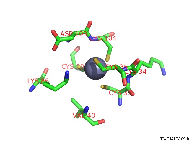
Mono view
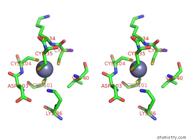
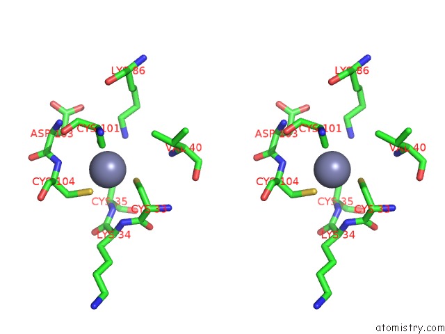
Stereo pair view
Mono view
Stereo pair view
A full contact list of Zinc with other atoms in the Zn binding
site number 1 of Crystal Structure Analysis of Glutathione-Dependent Formaldehyde- Activating Enzyme (Gfa) within 5.0Å range:
|
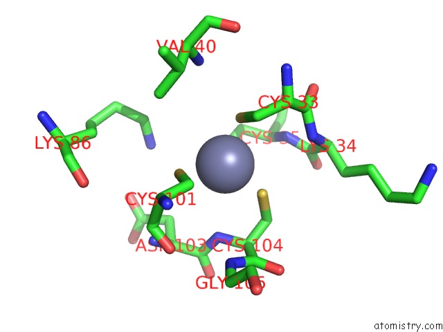
Zinc binding site 2 out of 4 in 1xa8
Go back to
Zinc binding site 2 out
of 4 in the Crystal Structure Analysis of Glutathione-Dependent Formaldehyde- Activating Enzyme (Gfa)
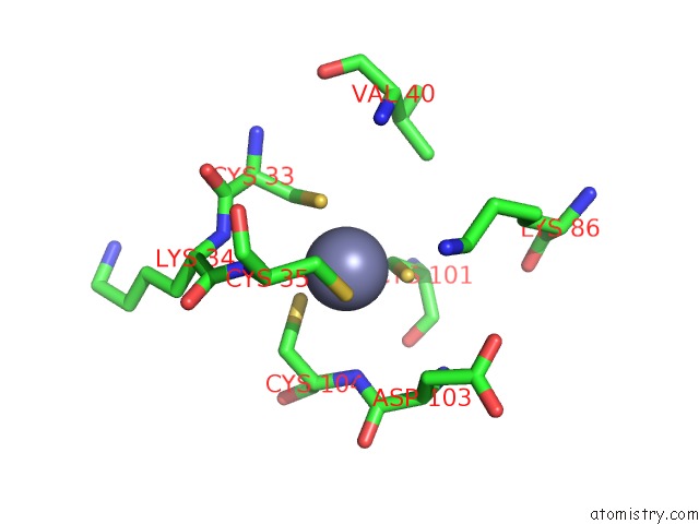
Mono view
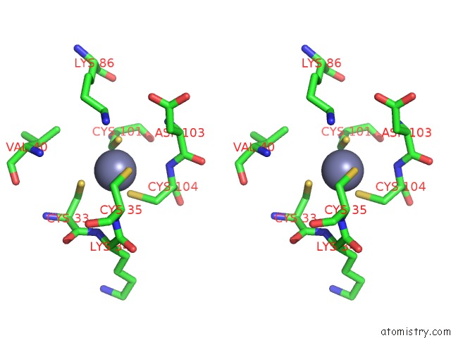
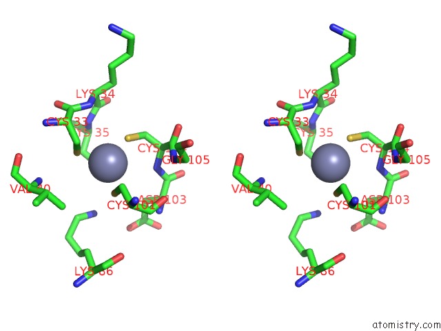
Stereo pair view
Mono view
Stereo pair view
A full contact list of Zinc with other atoms in the Zn binding
site number 2 of Crystal Structure Analysis of Glutathione-Dependent Formaldehyde- Activating Enzyme (Gfa) within 5.0Å range:
|
Zinc binding site 3 out of 4 in 1xa8
Go back to
Zinc binding site 3 out
of 4 in the Crystal Structure Analysis of Glutathione-Dependent Formaldehyde- Activating Enzyme (Gfa)
Mono view
Stereo pair view
Mono view
Stereo pair view
A full contact list of Zinc with other atoms in the Zn binding
site number 3 of Crystal Structure Analysis of Glutathione-Dependent Formaldehyde- Activating Enzyme (Gfa) within 5.0Å range:
|
Zinc binding site 4 out of 4 in 1xa8
Go back to
Zinc binding site 4 out
of 4 in the Crystal Structure Analysis of Glutathione-Dependent Formaldehyde- Activating Enzyme (Gfa)
Mono view
Stereo pair view
Mono view
Stereo pair view
A full contact list of Zinc with other atoms in the Zn binding
site number 4 of Crystal Structure Analysis of Glutathione-Dependent Formaldehyde- Activating Enzyme (Gfa) within 5.0Å range:
|
Reference:
A.M.Neculai,
D.Neculai,
C.Griesinger,
J.A.Vorholt,
S.Becker.
A Dynamic Zinc Redox Switch J.Biol.Chem. V. 280 2826 2005.
ISSN: ISSN 0021-9258
PubMed: 15548539
DOI: 10.1074/JBC.C400517200
Page generated: Wed Aug 20 00:13:26 2025
ISSN: ISSN 0021-9258
PubMed: 15548539
DOI: 10.1074/JBC.C400517200
Last articles
Zn in 2HAEZn in 2H6T
Zn in 2H6S
Zn in 2H6I
Zn in 2H6H
Zn in 2H6G
Zn in 2H6E
Zn in 2H6A
Zn in 2H6F
Zn in 2H59