Zinc »
PDB 2gzi-2han »
2h59 »
Zinc in PDB 2h59: SIR2 H116A-Deacetylated P53 Peptide-3'-O-Acetyl Adp Ribose
Protein crystallography data
The structure of SIR2 H116A-Deacetylated P53 Peptide-3'-O-Acetyl Adp Ribose, PDB code: 2h59
was solved by
K.G.Hoff,
J.L.Avalos,
K.Sens,
C.Wolberger,
with X-Ray Crystallography technique. A brief refinement statistics is given in the table below:
| Resolution Low / High (Å) | 50.00 / 1.90 |
| Space group | P 41 |
| Cell size a, b, c (Å), α, β, γ (°) | 46.996, 46.996, 257.081, 90.00, 90.00, 90.00 |
| R / Rfree (%) | 20.5 / 24.7 |
Zinc Binding Sites:
The binding sites of Zinc atom in the SIR2 H116A-Deacetylated P53 Peptide-3'-O-Acetyl Adp Ribose
(pdb code 2h59). This binding sites where shown within
5.0 Angstroms radius around Zinc atom.
In total 2 binding sites of Zinc where determined in the SIR2 H116A-Deacetylated P53 Peptide-3'-O-Acetyl Adp Ribose, PDB code: 2h59:
Jump to Zinc binding site number: 1; 2;
In total 2 binding sites of Zinc where determined in the SIR2 H116A-Deacetylated P53 Peptide-3'-O-Acetyl Adp Ribose, PDB code: 2h59:
Jump to Zinc binding site number: 1; 2;
Zinc binding site 1 out of 2 in 2h59
Go back to
Zinc binding site 1 out
of 2 in the SIR2 H116A-Deacetylated P53 Peptide-3'-O-Acetyl Adp Ribose
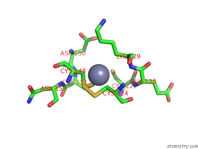
Mono view
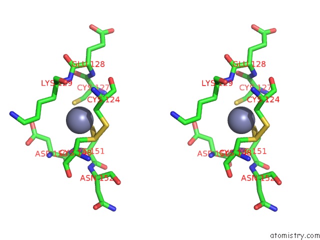
Stereo pair view
Mono view
Stereo pair view
A full contact list of Zinc with other atoms in the Zn binding
site number 1 of SIR2 H116A-Deacetylated P53 Peptide-3'-O-Acetyl Adp Ribose within 5.0Å range:
|
Zinc binding site 2 out of 2 in 2h59
Go back to
Zinc binding site 2 out
of 2 in the SIR2 H116A-Deacetylated P53 Peptide-3'-O-Acetyl Adp Ribose
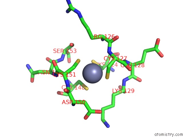
Mono view
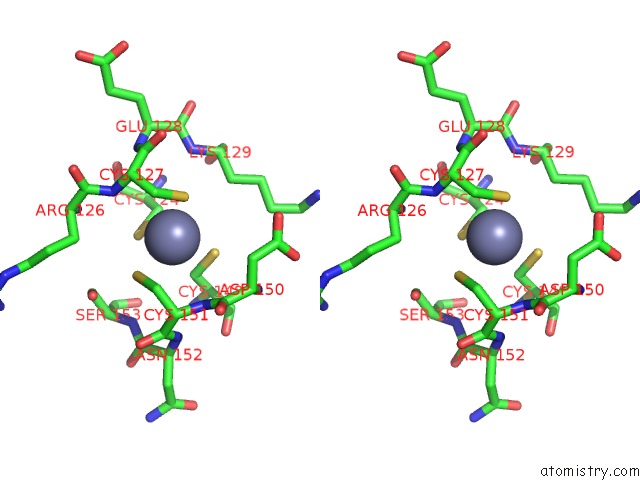
Stereo pair view
Mono view
Stereo pair view
A full contact list of Zinc with other atoms in the Zn binding
site number 2 of SIR2 H116A-Deacetylated P53 Peptide-3'-O-Acetyl Adp Ribose within 5.0Å range:
|
Reference:
K.G.Hoff,
J.L.Avalos,
K.Sens,
C.Wolberger.
Insights Into the Sirtuin Mechanism From Ternary Complexes Containing Nad(+) and Acetylated Peptide. Structure V. 14 1231 2006.
ISSN: ISSN 0969-2126
PubMed: 16905097
DOI: 10.1016/J.STR.2006.06.006
Page generated: Wed Aug 20 03:17:14 2025
ISSN: ISSN 0969-2126
PubMed: 16905097
DOI: 10.1016/J.STR.2006.06.006
Last articles
Zn in 3AV6Zn in 3AX1
Zn in 3AVS
Zn in 3AV5
Zn in 3AV4
Zn in 3AVR
Zn in 3AVE
Zn in 3AUO
Zn in 3AU2
Zn in 3AUK