Zinc »
PDB 3adr-3avs »
3av6 »
Zinc in PDB 3av6: Crystal Structure of Mouse Dna Methyltransferase 1 with Adomet
Enzymatic activity of Crystal Structure of Mouse Dna Methyltransferase 1 with Adomet
All present enzymatic activity of Crystal Structure of Mouse Dna Methyltransferase 1 with Adomet:
2.1.1.37;
2.1.1.37;
Protein crystallography data
The structure of Crystal Structure of Mouse Dna Methyltransferase 1 with Adomet, PDB code: 3av6
was solved by
K.Takeshita,
I.Suetake,
E.Yamashita,
M.Suga,
H.Narita,
A.Nakagawa,
S.Tajima,
with X-Ray Crystallography technique. A brief refinement statistics is given in the table below:
| Resolution Low / High (Å) | 43.14 / 3.09 |
| Space group | P 21 21 2 |
| Cell size a, b, c (Å), α, β, γ (°) | 134.995, 97.139, 130.794, 90.00, 90.00, 90.00 |
| R / Rfree (%) | 19.1 / 25.5 |
Zinc Binding Sites:
The binding sites of Zinc atom in the Crystal Structure of Mouse Dna Methyltransferase 1 with Adomet
(pdb code 3av6). This binding sites where shown within
5.0 Angstroms radius around Zinc atom.
In total 4 binding sites of Zinc where determined in the Crystal Structure of Mouse Dna Methyltransferase 1 with Adomet, PDB code: 3av6:
Jump to Zinc binding site number: 1; 2; 3; 4;
In total 4 binding sites of Zinc where determined in the Crystal Structure of Mouse Dna Methyltransferase 1 with Adomet, PDB code: 3av6:
Jump to Zinc binding site number: 1; 2; 3; 4;
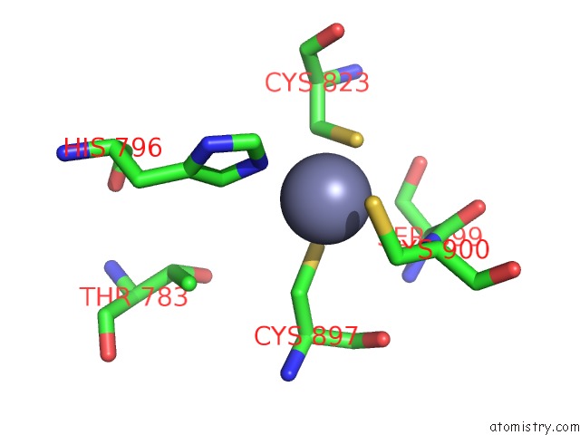
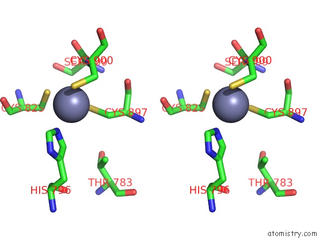
Zinc binding site 1 out of 4 in 3av6
Go back to
Zinc binding site 1 out
of 4 in the Crystal Structure of Mouse Dna Methyltransferase 1 with Adomet
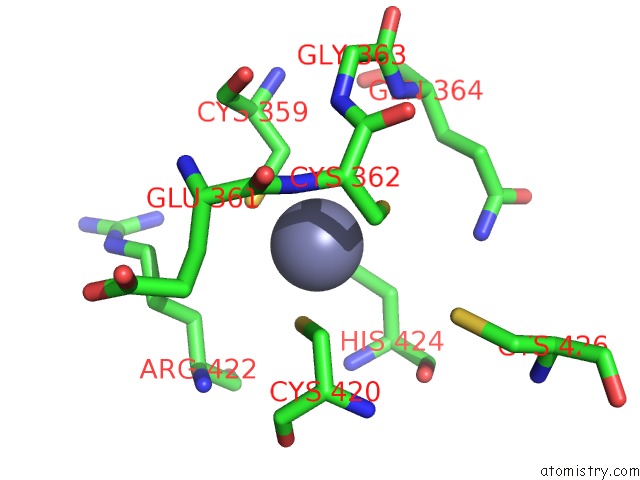
Mono view
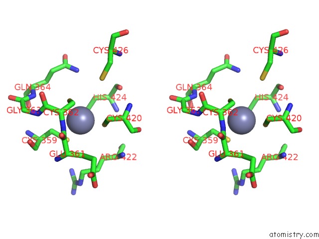
Stereo pair view
Mono view
Stereo pair view
A full contact list of Zinc with other atoms in the Zn binding
site number 1 of Crystal Structure of Mouse Dna Methyltransferase 1 with Adomet within 5.0Å range:
|
Zinc binding site 2 out of 4 in 3av6
Go back to
Zinc binding site 2 out
of 4 in the Crystal Structure of Mouse Dna Methyltransferase 1 with Adomet
Mono view
Stereo pair view
Mono view
Stereo pair view
A full contact list of Zinc with other atoms in the Zn binding
site number 2 of Crystal Structure of Mouse Dna Methyltransferase 1 with Adomet within 5.0Å range:
|
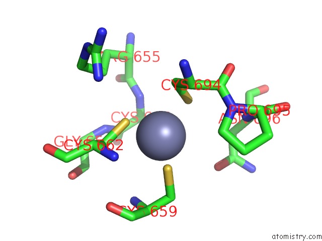
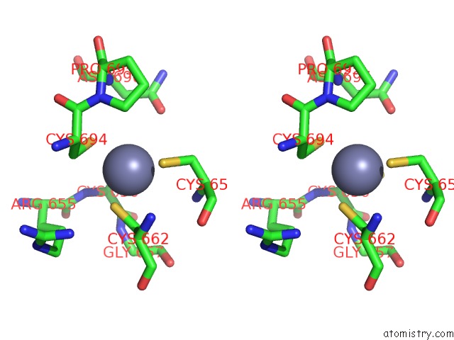
Zinc binding site 3 out of 4 in 3av6
Go back to
Zinc binding site 3 out
of 4 in the Crystal Structure of Mouse Dna Methyltransferase 1 with Adomet
Mono view
Stereo pair view
Mono view
Stereo pair view
A full contact list of Zinc with other atoms in the Zn binding
site number 3 of Crystal Structure of Mouse Dna Methyltransferase 1 with Adomet within 5.0Å range:
|
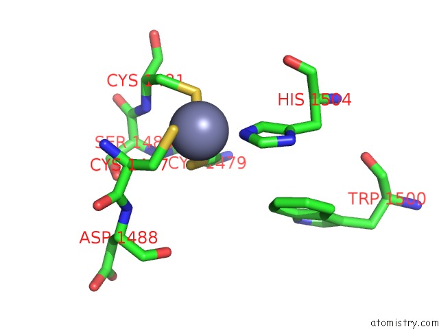
Zinc binding site 4 out of 4 in 3av6
Go back to
Zinc binding site 4 out
of 4 in the Crystal Structure of Mouse Dna Methyltransferase 1 with Adomet
Mono view
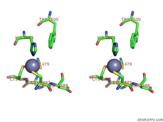
Stereo pair view
Mono view
Stereo pair view
A full contact list of Zinc with other atoms in the Zn binding
site number 4 of Crystal Structure of Mouse Dna Methyltransferase 1 with Adomet within 5.0Å range:
|
Reference:
K.Takeshita,
I.Suetake,
E.Yamashita,
M.Suga,
H.Narita,
A.Nakagawa,
S.Tajima.
Structural Insight Into Maintenance Methylation By Mouse Dna Methyltransferase 1 (DNMT1). Proc.Natl.Acad.Sci.Usa V. 108 9055 2011.
ISSN: ESSN 1091-6490
PubMed: 21518897
DOI: 10.1073/PNAS.1019629108
Page generated: Thu Oct 24 11:17:35 2024
ISSN: ESSN 1091-6490
PubMed: 21518897
DOI: 10.1073/PNAS.1019629108
Last articles
Mg in 7EUQMg in 7EUN
Mg in 7EV3
Mg in 7EV2
Mg in 7EUL
Mg in 7EUK
Mg in 7EU1
Mg in 7ETE
Mg in 7EU0
Mg in 7ESQ