Zinc »
PDB 1v0d-1vdd »
1v9e »
Zinc in PDB 1v9e: Crystal Structure Analysis of Bovine Carbonic Anhydrase II
Enzymatic activity of Crystal Structure Analysis of Bovine Carbonic Anhydrase II
All present enzymatic activity of Crystal Structure Analysis of Bovine Carbonic Anhydrase II:
4.2.1.1;
4.2.1.1;
Protein crystallography data
The structure of Crystal Structure Analysis of Bovine Carbonic Anhydrase II, PDB code: 1v9e
was solved by
R.Saito,
T.Sato,
A.Ikai,
N.Tanaka,
with X-Ray Crystallography technique. A brief refinement statistics is given in the table below:
| Resolution Low / High (Å) | 17.87 / 1.95 |
| Space group | P 61 |
| Cell size a, b, c (Å), α, β, γ (°) | 66.700, 66.700, 240.000, 90.00, 90.00, 120.00 |
| R / Rfree (%) | 27 / 29.8 |
Zinc Binding Sites:
The binding sites of Zinc atom in the Crystal Structure Analysis of Bovine Carbonic Anhydrase II
(pdb code 1v9e). This binding sites where shown within
5.0 Angstroms radius around Zinc atom.
In total 2 binding sites of Zinc where determined in the Crystal Structure Analysis of Bovine Carbonic Anhydrase II, PDB code: 1v9e:
Jump to Zinc binding site number: 1; 2;
In total 2 binding sites of Zinc where determined in the Crystal Structure Analysis of Bovine Carbonic Anhydrase II, PDB code: 1v9e:
Jump to Zinc binding site number: 1; 2;
Zinc binding site 1 out of 2 in 1v9e
Go back to
Zinc binding site 1 out
of 2 in the Crystal Structure Analysis of Bovine Carbonic Anhydrase II
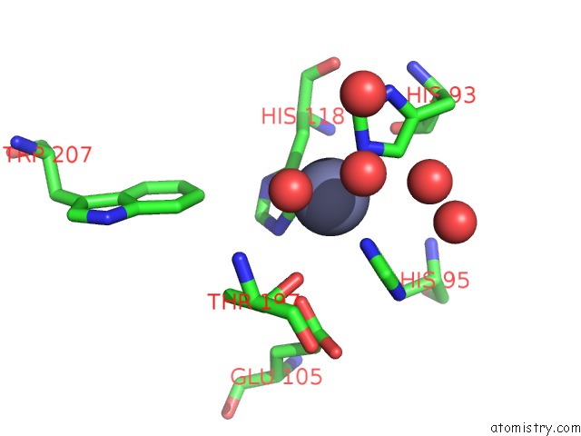
Mono view
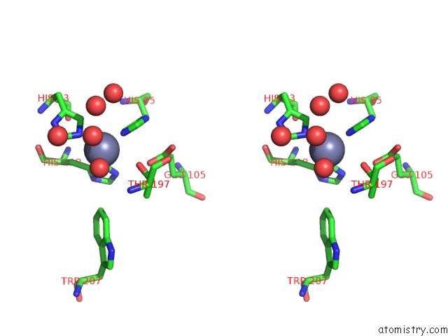
Stereo pair view
Mono view
Stereo pair view
A full contact list of Zinc with other atoms in the Zn binding
site number 1 of Crystal Structure Analysis of Bovine Carbonic Anhydrase II within 5.0Å range:
|
Zinc binding site 2 out of 2 in 1v9e
Go back to
Zinc binding site 2 out
of 2 in the Crystal Structure Analysis of Bovine Carbonic Anhydrase II
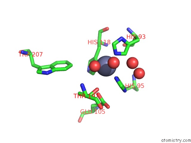
Mono view
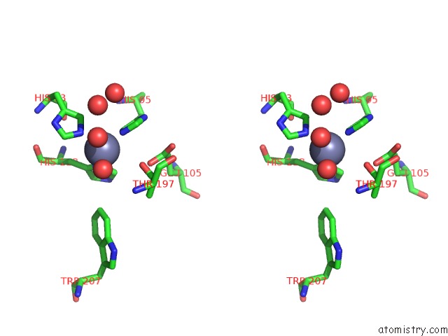
Stereo pair view
Mono view
Stereo pair view
A full contact list of Zinc with other atoms in the Zn binding
site number 2 of Crystal Structure Analysis of Bovine Carbonic Anhydrase II within 5.0Å range:
|
Reference:
R.Saito,
T.Sato,
A.Ikai,
N.Tanaka.
Structure of Bovine Carbonic Anhydrase II at 1.95 A Resolution. Acta Crystallogr.,Sect.D V. 60 792 2004.
ISSN: ISSN 0907-4449
PubMed: 15039588
DOI: 10.1107/S0907444904003166
Page generated: Wed Oct 16 19:46:25 2024
ISSN: ISSN 0907-4449
PubMed: 15039588
DOI: 10.1107/S0907444904003166
Last articles
Zn in 9JPJZn in 9JP7
Zn in 9JPK
Zn in 9JPL
Zn in 9GN6
Zn in 9GN7
Zn in 9GKU
Zn in 9GKW
Zn in 9GKX
Zn in 9GL0