Zinc »
PDB 1v0d-1vdd »
1v33 »
Zinc in PDB 1v33: Crystal Structure of Dna Primase From Pyrococcus Horikoshii
Protein crystallography data
The structure of Crystal Structure of Dna Primase From Pyrococcus Horikoshii, PDB code: 1v33
was solved by
N.Ito,
O.Nureki,
M.Shirouzu,
S.Yokoyama,
F.Hanaoka,
Rikenstructural Genomics/Proteomics Initiative (Rsgi),
with X-Ray Crystallography technique. A brief refinement statistics is given in the table below:
| Resolution Low / High (Å) | 33.10 / 1.80 |
| Space group | P 32 2 1 |
| Cell size a, b, c (Å), α, β, γ (°) | 76.451, 76.451, 128.894, 90.00, 90.00, 120.00 |
| R / Rfree (%) | 21.9 / 24.4 |
Zinc Binding Sites:
The binding sites of Zinc atom in the Crystal Structure of Dna Primase From Pyrococcus Horikoshii
(pdb code 1v33). This binding sites where shown within
5.0 Angstroms radius around Zinc atom.
In total only one binding site of Zinc was determined in the Crystal Structure of Dna Primase From Pyrococcus Horikoshii, PDB code: 1v33:
In total only one binding site of Zinc was determined in the Crystal Structure of Dna Primase From Pyrococcus Horikoshii, PDB code: 1v33:
Zinc binding site 1 out of 1 in 1v33
Go back to
Zinc binding site 1 out
of 1 in the Crystal Structure of Dna Primase From Pyrococcus Horikoshii
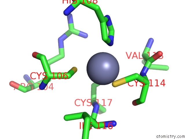
Mono view
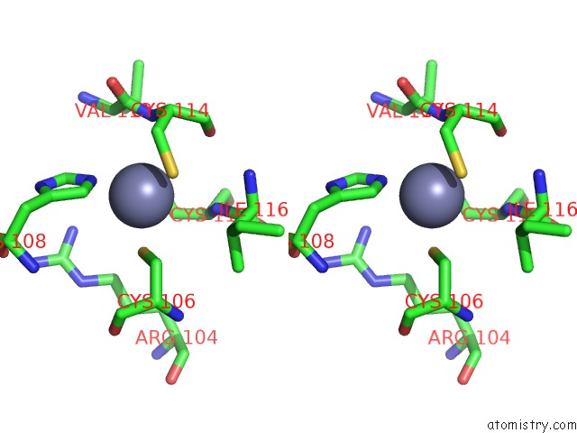
Stereo pair view
Mono view
Stereo pair view
A full contact list of Zinc with other atoms in the Zn binding
site number 1 of Crystal Structure of Dna Primase From Pyrococcus Horikoshii within 5.0Å range:
|
Reference:
N.Ito,
O.Nureki,
M.Shirouzu,
S.Yokoyama,
F.Hanaoka.
Crystal Structure of the Pyrococcus Horikoshii Dna Primase-Utp Complex: Implications For the Mechanism of Primer Synthesis. Genes Cells V. 8 913 2003.
ISSN: ISSN 1356-9597
PubMed: 14750947
DOI: 10.1111/J.1365-2443.2003.00693.X
Page generated: Wed Oct 16 19:41:55 2024
ISSN: ISSN 1356-9597
PubMed: 14750947
DOI: 10.1111/J.1365-2443.2003.00693.X
Last articles
Zn in 9JPJZn in 9JP7
Zn in 9JPK
Zn in 9JPL
Zn in 9GN6
Zn in 9GN7
Zn in 9GKU
Zn in 9GKW
Zn in 9GKX
Zn in 9GL0