Zinc »
PDB 1my2-1ndy »
1ndw »
Zinc in PDB 1ndw: Crystal Structure of Adenosine Deaminase Complexed with FR221647
Enzymatic activity of Crystal Structure of Adenosine Deaminase Complexed with FR221647
All present enzymatic activity of Crystal Structure of Adenosine Deaminase Complexed with FR221647:
3.5.4.4;
3.5.4.4;
Protein crystallography data
The structure of Crystal Structure of Adenosine Deaminase Complexed with FR221647, PDB code: 1ndw
was solved by
T.Kinoshita,
with X-Ray Crystallography technique. A brief refinement statistics is given in the table below:
| Resolution Low / High (Å) | 8.00 / 2.00 |
| Space group | P 43 21 2 |
| Cell size a, b, c (Å), α, β, γ (°) | 77.630, 77.630, 135.660, 90.00, 90.00, 90.00 |
| R / Rfree (%) | n/a / n/a |
Zinc Binding Sites:
The binding sites of Zinc atom in the Crystal Structure of Adenosine Deaminase Complexed with FR221647
(pdb code 1ndw). This binding sites where shown within
5.0 Angstroms radius around Zinc atom.
In total only one binding site of Zinc was determined in the Crystal Structure of Adenosine Deaminase Complexed with FR221647, PDB code: 1ndw:
In total only one binding site of Zinc was determined in the Crystal Structure of Adenosine Deaminase Complexed with FR221647, PDB code: 1ndw:
Zinc binding site 1 out of 1 in 1ndw
Go back to
Zinc binding site 1 out
of 1 in the Crystal Structure of Adenosine Deaminase Complexed with FR221647
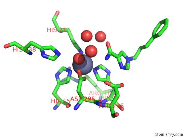
Mono view
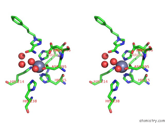
Stereo pair view
Mono view
Stereo pair view
A full contact list of Zinc with other atoms in the Zn binding
site number 1 of Crystal Structure of Adenosine Deaminase Complexed with FR221647 within 5.0Å range:
|
Reference:
T.Terasaka,
T.Kinoshita,
M.Kuno,
I.Nakanishi.
A Highly Potent Non-Nucleoside Adenosine Deaminase Inhibitor: Efficient Drug Discovery By Intentional Lead Hybridization J.Am.Chem.Soc. V. 126 34 2004.
ISSN: ISSN 0002-7863
PubMed: 14709046
DOI: 10.1021/JA038606L
Page generated: Tue Aug 19 21:57:26 2025
ISSN: ISSN 0002-7863
PubMed: 14709046
DOI: 10.1021/JA038606L
Last articles
Zn in 1XTGZn in 1XSO
Zn in 1XRY
Zn in 1XRZ
Zn in 1XRT
Zn in 1XRU
Zn in 1XRF
Zn in 1XQ0
Zn in 1XOC
Zn in 1XPZ