Zinc »
PDB 1xmo-1xur »
1xso »
Zinc in PDB 1xso: Three-Dimensional Structure of Xenopus Laevis Cu,Zn Superoxide Dismutase B Determined By X-Ray Crystallography at 1.5 Angstroms Resolution
Enzymatic activity of Three-Dimensional Structure of Xenopus Laevis Cu,Zn Superoxide Dismutase B Determined By X-Ray Crystallography at 1.5 Angstroms Resolution
All present enzymatic activity of Three-Dimensional Structure of Xenopus Laevis Cu,Zn Superoxide Dismutase B Determined By X-Ray Crystallography at 1.5 Angstroms Resolution:
1.15.1.1;
1.15.1.1;
Protein crystallography data
The structure of Three-Dimensional Structure of Xenopus Laevis Cu,Zn Superoxide Dismutase B Determined By X-Ray Crystallography at 1.5 Angstroms Resolution, PDB code: 1xso
was solved by
K.Djinovic Carugo,
A.Coda,
A.Battistoni,
M.T.Carri,
F.Polticelli,
A.Desideri,
G.Rotilio,
K.S.Wilson,
M.Bolognesi,
with X-Ray Crystallography technique. A brief refinement statistics is given in the table below:
| Resolution Low / High (Å) | 10.00 / 1.49 |
| Space group | P 21 21 21 |
| Cell size a, b, c (Å), α, β, γ (°) | 73.450, 68.940, 58.760, 90.00, 90.00, 90.00 |
| R / Rfree (%) | 10.4 / 16.9 |
Other elements in 1xso:
The structure of Three-Dimensional Structure of Xenopus Laevis Cu,Zn Superoxide Dismutase B Determined By X-Ray Crystallography at 1.5 Angstroms Resolution also contains other interesting chemical elements:
| Copper | (Cu) | 2 atoms |
Zinc Binding Sites:
The binding sites of Zinc atom in the Three-Dimensional Structure of Xenopus Laevis Cu,Zn Superoxide Dismutase B Determined By X-Ray Crystallography at 1.5 Angstroms Resolution
(pdb code 1xso). This binding sites where shown within
5.0 Angstroms radius around Zinc atom.
In total 2 binding sites of Zinc where determined in the Three-Dimensional Structure of Xenopus Laevis Cu,Zn Superoxide Dismutase B Determined By X-Ray Crystallography at 1.5 Angstroms Resolution, PDB code: 1xso:
Jump to Zinc binding site number: 1; 2;
In total 2 binding sites of Zinc where determined in the Three-Dimensional Structure of Xenopus Laevis Cu,Zn Superoxide Dismutase B Determined By X-Ray Crystallography at 1.5 Angstroms Resolution, PDB code: 1xso:
Jump to Zinc binding site number: 1; 2;
Zinc binding site 1 out of 2 in 1xso
Go back to
Zinc binding site 1 out
of 2 in the Three-Dimensional Structure of Xenopus Laevis Cu,Zn Superoxide Dismutase B Determined By X-Ray Crystallography at 1.5 Angstroms Resolution
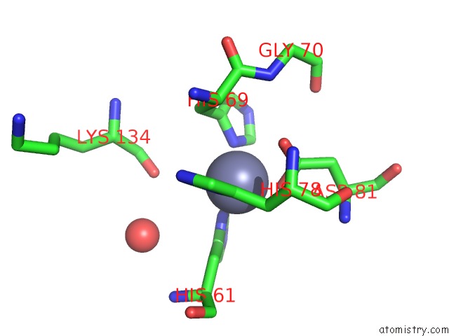
Mono view
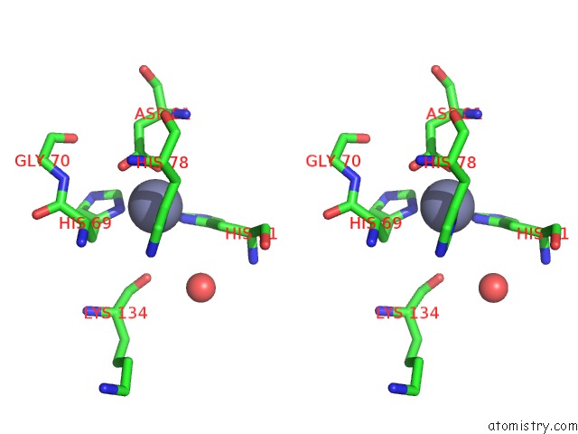
Stereo pair view
Mono view
Stereo pair view
A full contact list of Zinc with other atoms in the Zn binding
site number 1 of Three-Dimensional Structure of Xenopus Laevis Cu,Zn Superoxide Dismutase B Determined By X-Ray Crystallography at 1.5 Angstroms Resolution within 5.0Å range:
|
Zinc binding site 2 out of 2 in 1xso
Go back to
Zinc binding site 2 out
of 2 in the Three-Dimensional Structure of Xenopus Laevis Cu,Zn Superoxide Dismutase B Determined By X-Ray Crystallography at 1.5 Angstroms Resolution
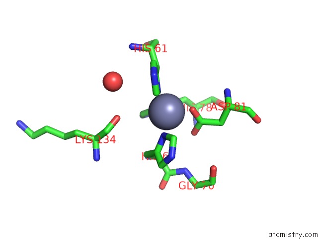
Mono view
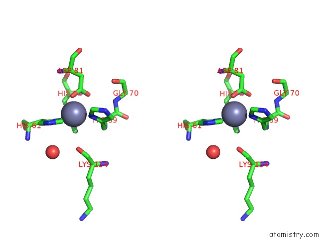
Stereo pair view
Mono view
Stereo pair view
A full contact list of Zinc with other atoms in the Zn binding
site number 2 of Three-Dimensional Structure of Xenopus Laevis Cu,Zn Superoxide Dismutase B Determined By X-Ray Crystallography at 1.5 Angstroms Resolution within 5.0Å range:
|
Reference:
K.Djinovic Carugo,
A.Battistoni,
M.T.Carri,
F.Polticelli,
A.Desideri,
G.Rotilio,
A.Coda,
K.S.Wilson,
M.Bolognesi.
Three-Dimensional Structure of Xenopus Laevis Cu,Zn Superoxide Dismutase B Determined By X-Ray Crystallography at 1.5 A Resolution. Acta Crystallogr.,Sect.D V. 52 176 1996.
ISSN: ISSN 0907-4449
PubMed: 15299740
DOI: 10.1107/S0907444995007608
Page generated: Wed Oct 16 20:35:14 2024
ISSN: ISSN 0907-4449
PubMed: 15299740
DOI: 10.1107/S0907444995007608
Last articles
I in 6UN0I in 6UMY
I in 6UKF
I in 6UKE
I in 6U2D
I in 6U9B
I in 6U9A
I in 6U8H
I in 6U98
I in 6U99