Zinc »
PDB 1xmo-1xur »
1xtg »
Zinc in PDB 1xtg: Crystal Structure of Neurotoxin Bont/A Complexed with Synaptosomal- Associated Protein 25
Protein crystallography data
The structure of Crystal Structure of Neurotoxin Bont/A Complexed with Synaptosomal- Associated Protein 25, PDB code: 1xtg
was solved by
M.A.Breidenbach,
A.T.Brunger,
with X-Ray Crystallography technique. A brief refinement statistics is given in the table below:
| Resolution Low / High (Å) | 29.92 / 2.10 |
| Space group | P 43 21 2 |
| Cell size a, b, c (Å), α, β, γ (°) | 86.000, 86.000, 165.400, 90.00, 90.00, 90.00 |
| R / Rfree (%) | 21.8 / 24.7 |
Other elements in 1xtg:
The structure of Crystal Structure of Neurotoxin Bont/A Complexed with Synaptosomal- Associated Protein 25 also contains other interesting chemical elements:
| Chlorine | (Cl) | 1 atom |
Zinc Binding Sites:
The binding sites of Zinc atom in the Crystal Structure of Neurotoxin Bont/A Complexed with Synaptosomal- Associated Protein 25
(pdb code 1xtg). This binding sites where shown within
5.0 Angstroms radius around Zinc atom.
In total only one binding site of Zinc was determined in the Crystal Structure of Neurotoxin Bont/A Complexed with Synaptosomal- Associated Protein 25, PDB code: 1xtg:
In total only one binding site of Zinc was determined in the Crystal Structure of Neurotoxin Bont/A Complexed with Synaptosomal- Associated Protein 25, PDB code: 1xtg:
Zinc binding site 1 out of 1 in 1xtg
Go back to
Zinc binding site 1 out
of 1 in the Crystal Structure of Neurotoxin Bont/A Complexed with Synaptosomal- Associated Protein 25

Mono view
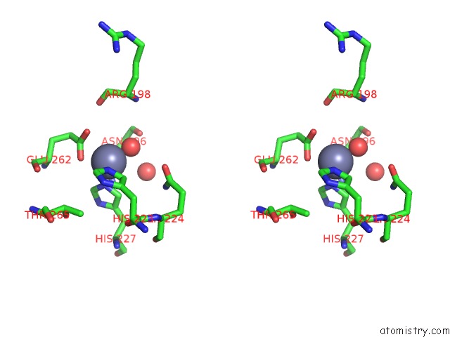
Stereo pair view
Mono view
Stereo pair view
A full contact list of Zinc with other atoms in the Zn binding
site number 1 of Crystal Structure of Neurotoxin Bont/A Complexed with Synaptosomal- Associated Protein 25 within 5.0Å range:
|
Reference:
M.A.Breidenbach,
A.T.Brunger.
Substrate Recognition Strategy For Botulinum Neurotoxin Serotype A Nature V. 432 925 2004.
ISSN: ISSN 0028-0836
PubMed: 15592454
DOI: 10.1038/NATURE03123
Page generated: Wed Oct 16 20:35:36 2024
ISSN: ISSN 0028-0836
PubMed: 15592454
DOI: 10.1038/NATURE03123
Last articles
Hg in 6RNPHg in 6RL9
Hg in 6RKN
Hg in 6RMP
Hg in 6RM1
Hg in 6IUE
Hg in 6RJJ
Hg in 6RIT
Hg in 6RIG
Hg in 6NWH