Zinc »
PDB 1kzy-1lg6 »
1lg5 »
Zinc in PDB 1lg5: Crystal Structure Analysis of the Hca II Mutant T199P in Complex with Beta-Mercaptoethanol
Enzymatic activity of Crystal Structure Analysis of the Hca II Mutant T199P in Complex with Beta-Mercaptoethanol
All present enzymatic activity of Crystal Structure Analysis of the Hca II Mutant T199P in Complex with Beta-Mercaptoethanol:
4.2.1.1;
4.2.1.1;
Protein crystallography data
The structure of Crystal Structure Analysis of the Hca II Mutant T199P in Complex with Beta-Mercaptoethanol, PDB code: 1lg5
was solved by
S.Huang,
B.Sjoblom,
A.E.Sauer-Eriksson,
B.-H.Jonsson,
with X-Ray Crystallography technique. A brief refinement statistics is given in the table below:
| Resolution Low / High (Å) | 20.00 / 1.75 |
| Space group | C 1 2 1 |
| Cell size a, b, c (Å), α, β, γ (°) | 72.840, 44.803, 76.457, 90.00, 92.51, 90.00 |
| R / Rfree (%) | 20.5 / 22.7 |
Zinc Binding Sites:
The binding sites of Zinc atom in the Crystal Structure Analysis of the Hca II Mutant T199P in Complex with Beta-Mercaptoethanol
(pdb code 1lg5). This binding sites where shown within
5.0 Angstroms radius around Zinc atom.
In total only one binding site of Zinc was determined in the Crystal Structure Analysis of the Hca II Mutant T199P in Complex with Beta-Mercaptoethanol, PDB code: 1lg5:
In total only one binding site of Zinc was determined in the Crystal Structure Analysis of the Hca II Mutant T199P in Complex with Beta-Mercaptoethanol, PDB code: 1lg5:
Zinc binding site 1 out of 1 in 1lg5
Go back to
Zinc binding site 1 out
of 1 in the Crystal Structure Analysis of the Hca II Mutant T199P in Complex with Beta-Mercaptoethanol
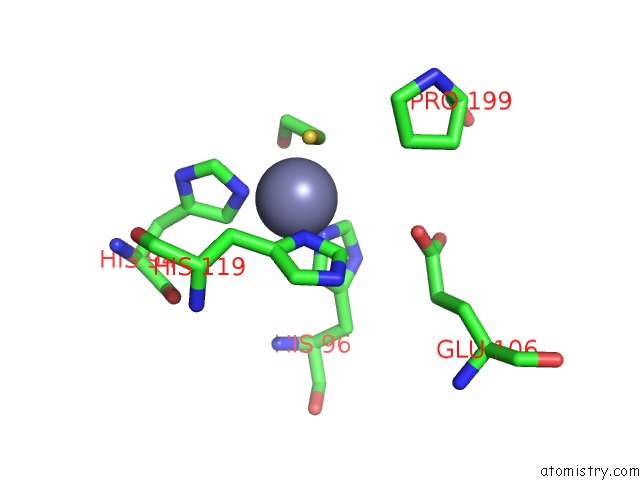
Mono view
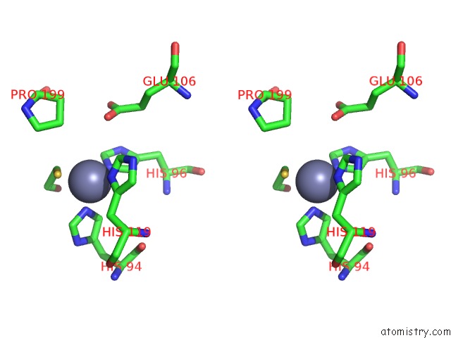
Stereo pair view
Mono view
Stereo pair view
A full contact list of Zinc with other atoms in the Zn binding
site number 1 of Crystal Structure Analysis of the Hca II Mutant T199P in Complex with Beta-Mercaptoethanol within 5.0Å range:
|
Reference:
S.Huang,
B.Sjoblom,
A.E.Sauer-Eriksson,
B.H.Jonsson.
Organization of An Efficient Carbonic Anhydrase: Implications For the Mechanism Based on Structure-Function Studies of A T199P/C206S Mutant. Biochemistry V. 41 7628 2002.
ISSN: ISSN 0006-2960
PubMed: 12056894
DOI: 10.1021/BI020053O
Page generated: Tue Aug 19 21:32:32 2025
ISSN: ISSN 0006-2960
PubMed: 12056894
DOI: 10.1021/BI020053O
Last articles
Zn in 1X4WZn in 1X4V
Zn in 1X4U
Zn in 1X4S
Zn in 1X4L
Zn in 1X4K
Zn in 1X4J
Zn in 1X4I
Zn in 1X3H
Zn in 1X3Z