Zinc »
PDB 1kzy-1lg6 »
1lfw »
Zinc in PDB 1lfw: Crystal Structure of Pepv
Enzymatic activity of Crystal Structure of Pepv
All present enzymatic activity of Crystal Structure of Pepv:
3.4.13.3;
3.4.13.3;
Protein crystallography data
The structure of Crystal Structure of Pepv, PDB code: 1lfw
was solved by
D.Jozic,
G.Bourenkow,
H.Bartunik,
H.Scholze,
V.Dive,
B.Henrich,
R.Huber,
W.Bode,
K.Maskos,
with X-Ray Crystallography technique. A brief refinement statistics is given in the table below:
| Resolution Low / High (Å) | 11.00 / 1.80 |
| Space group | P 21 21 21 |
| Cell size a, b, c (Å), α, β, γ (°) | 67.151, 77.025, 89.955, 90.00, 90.00, 90.00 |
| R / Rfree (%) | 17.4 / n/a |
Zinc Binding Sites:
The binding sites of Zinc atom in the Crystal Structure of Pepv
(pdb code 1lfw). This binding sites where shown within
5.0 Angstroms radius around Zinc atom.
In total 2 binding sites of Zinc where determined in the Crystal Structure of Pepv, PDB code: 1lfw:
Jump to Zinc binding site number: 1; 2;
In total 2 binding sites of Zinc where determined in the Crystal Structure of Pepv, PDB code: 1lfw:
Jump to Zinc binding site number: 1; 2;
Zinc binding site 1 out of 2 in 1lfw
Go back to
Zinc binding site 1 out
of 2 in the Crystal Structure of Pepv
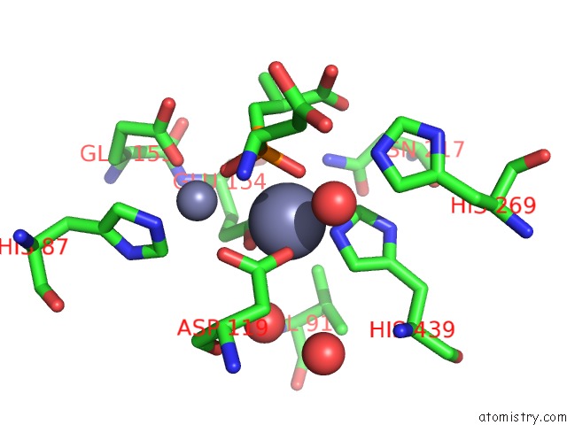
Mono view
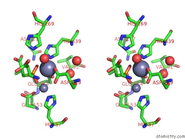
Stereo pair view
Mono view
Stereo pair view
A full contact list of Zinc with other atoms in the Zn binding
site number 1 of Crystal Structure of Pepv within 5.0Å range:
|
Zinc binding site 2 out of 2 in 1lfw
Go back to
Zinc binding site 2 out
of 2 in the Crystal Structure of Pepv
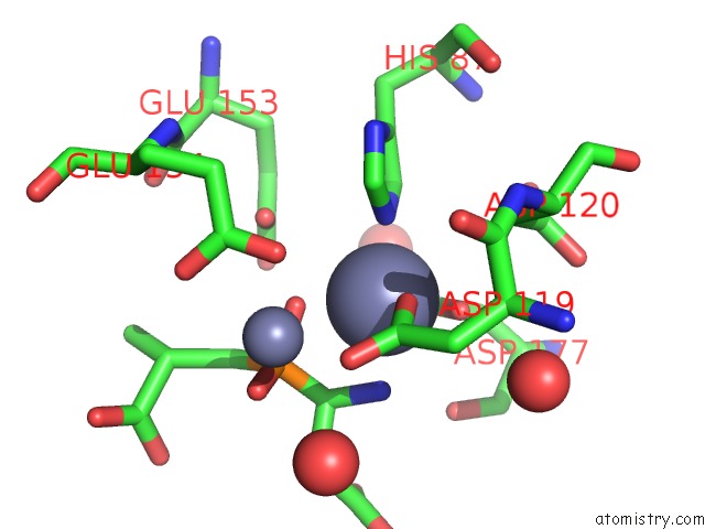
Mono view
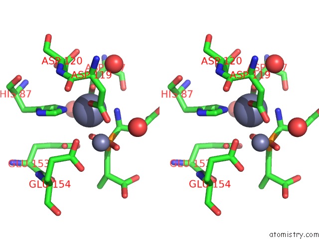
Stereo pair view
Mono view
Stereo pair view
A full contact list of Zinc with other atoms in the Zn binding
site number 2 of Crystal Structure of Pepv within 5.0Å range:
|
Reference:
D.Jozic,
G.Bourenkow,
H.Bartunik,
H.Scholze,
V.Dive,
B.Henrich,
R.Huber,
W.Bode,
K.Maskos.
Crystal Structure of the Dinuclear Zinc Aminopeptidase Pepv From Lactobacillus Delbrueckii Unravels Its Preference For Dipeptides Structure V. 10 1097 2002.
ISSN: ISSN 0969-2126
PubMed: 12176387
DOI: 10.1016/S0969-2126(02)00805-5
Page generated: Tue Aug 19 21:32:21 2025
ISSN: ISSN 0969-2126
PubMed: 12176387
DOI: 10.1016/S0969-2126(02)00805-5
Last articles
Zn in 1X4KZn in 1X4J
Zn in 1X4I
Zn in 1X3H
Zn in 1X3Z
Zn in 1X3W
Zn in 1X3C
Zn in 1X1C
Zn in 1X1V
Zn in 1X31