Zinc »
PDB 1cp6-1d4u »
1d1v »
Zinc in PDB 1d1v: Bovine Endothelial Nitric Oxide Synthase Heme Domain Complexed with S- Ethyl-N-Phenyl-Isothiourea (H4B Bound)
Enzymatic activity of Bovine Endothelial Nitric Oxide Synthase Heme Domain Complexed with S- Ethyl-N-Phenyl-Isothiourea (H4B Bound)
All present enzymatic activity of Bovine Endothelial Nitric Oxide Synthase Heme Domain Complexed with S- Ethyl-N-Phenyl-Isothiourea (H4B Bound):
1.14.13.39;
1.14.13.39;
Protein crystallography data
The structure of Bovine Endothelial Nitric Oxide Synthase Heme Domain Complexed with S- Ethyl-N-Phenyl-Isothiourea (H4B Bound), PDB code: 1d1v
was solved by
C.S.Raman,
H.Li,
P.Martasek,
G.J.Southan,
B.S.S.Masters,
T.L.Poulos,
with X-Ray Crystallography technique. A brief refinement statistics is given in the table below:
| Resolution Low / High (Å) | 36.48 / 1.93 |
| Space group | P 21 21 21 |
| Cell size a, b, c (Å), α, β, γ (°) | 57.940, 106.560, 156.950, 90.00, 90.00, 90.00 |
| R / Rfree (%) | 18.6 / 21.9 |
Other elements in 1d1v:
The structure of Bovine Endothelial Nitric Oxide Synthase Heme Domain Complexed with S- Ethyl-N-Phenyl-Isothiourea (H4B Bound) also contains other interesting chemical elements:
| Arsenic | (As) | 2 atoms |
| Iron | (Fe) | 2 atoms |
Zinc Binding Sites:
The binding sites of Zinc atom in the Bovine Endothelial Nitric Oxide Synthase Heme Domain Complexed with S- Ethyl-N-Phenyl-Isothiourea (H4B Bound)
(pdb code 1d1v). This binding sites where shown within
5.0 Angstroms radius around Zinc atom.
In total only one binding site of Zinc was determined in the Bovine Endothelial Nitric Oxide Synthase Heme Domain Complexed with S- Ethyl-N-Phenyl-Isothiourea (H4B Bound), PDB code: 1d1v:
In total only one binding site of Zinc was determined in the Bovine Endothelial Nitric Oxide Synthase Heme Domain Complexed with S- Ethyl-N-Phenyl-Isothiourea (H4B Bound), PDB code: 1d1v:
Zinc binding site 1 out of 1 in 1d1v
Go back to
Zinc binding site 1 out
of 1 in the Bovine Endothelial Nitric Oxide Synthase Heme Domain Complexed with S- Ethyl-N-Phenyl-Isothiourea (H4B Bound)
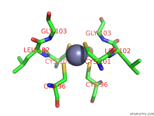
Mono view
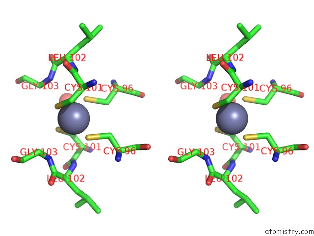
Stereo pair view
Mono view
Stereo pair view
A full contact list of Zinc with other atoms in the Zn binding
site number 1 of Bovine Endothelial Nitric Oxide Synthase Heme Domain Complexed with S- Ethyl-N-Phenyl-Isothiourea (H4B Bound) within 5.0Å range:
|
Reference:
C.S.Raman,
H.Li,
P.Martasek,
B.R.Babu,
O.W.Griffith,
B.S.Masters,
T.L.Poulos.
Implications For Isoform-Selective Inhibitor Design Derived From the Binding Mode of Bulky Isothioureas to the Heme Domain of Endothelial Nitric-Oxide Synthase. J.Biol.Chem. V. 276 26486 2001.
ISSN: ISSN 0021-9258
PubMed: 11331290
DOI: 10.1074/JBC.M102255200
Page generated: Sat Oct 12 23:27:00 2024
ISSN: ISSN 0021-9258
PubMed: 11331290
DOI: 10.1074/JBC.M102255200
Last articles
Zn in 9JPJZn in 9JP7
Zn in 9JPK
Zn in 9JPL
Zn in 9GN6
Zn in 9GN7
Zn in 9GKU
Zn in 9GKW
Zn in 9GKX
Zn in 9GL0