Zinc »
PDB 3wok-3x17 »
3wv6 »
Zinc in PDB 3wv6: Crystal Structure of A Protease-Resistant Mutant Form of Human Galectin-9
Protein crystallography data
The structure of Crystal Structure of A Protease-Resistant Mutant Form of Human Galectin-9, PDB code: 3wv6
was solved by
H.Yoshida,
S.Kamitori,
with X-Ray Crystallography technique. A brief refinement statistics is given in the table below:
| Resolution Low / High (Å) | 27.85 / 1.95 |
| Space group | P 1 21 1 |
| Cell size a, b, c (Å), α, β, γ (°) | 58.340, 94.870, 64.810, 90.00, 113.87, 90.00 |
| R / Rfree (%) | 18.8 / 22.2 |
Zinc Binding Sites:
The binding sites of Zinc atom in the Crystal Structure of A Protease-Resistant Mutant Form of Human Galectin-9
(pdb code 3wv6). This binding sites where shown within
5.0 Angstroms radius around Zinc atom.
In total 2 binding sites of Zinc where determined in the Crystal Structure of A Protease-Resistant Mutant Form of Human Galectin-9, PDB code: 3wv6:
Jump to Zinc binding site number: 1; 2;
In total 2 binding sites of Zinc where determined in the Crystal Structure of A Protease-Resistant Mutant Form of Human Galectin-9, PDB code: 3wv6:
Jump to Zinc binding site number: 1; 2;
Zinc binding site 1 out of 2 in 3wv6
Go back to
Zinc binding site 1 out
of 2 in the Crystal Structure of A Protease-Resistant Mutant Form of Human Galectin-9
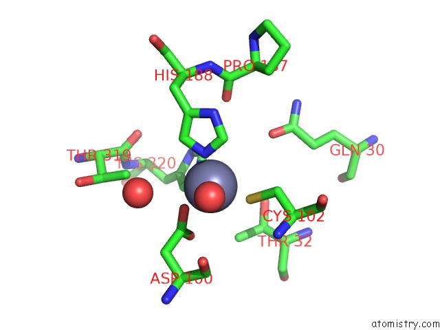
Mono view
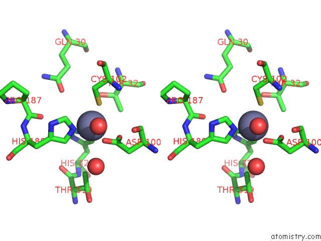
Stereo pair view
Mono view
Stereo pair view
A full contact list of Zinc with other atoms in the Zn binding
site number 1 of Crystal Structure of A Protease-Resistant Mutant Form of Human Galectin-9 within 5.0Å range:
|
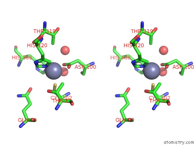
Zinc binding site 2 out of 2 in 3wv6
Go back to
Zinc binding site 2 out
of 2 in the Crystal Structure of A Protease-Resistant Mutant Form of Human Galectin-9
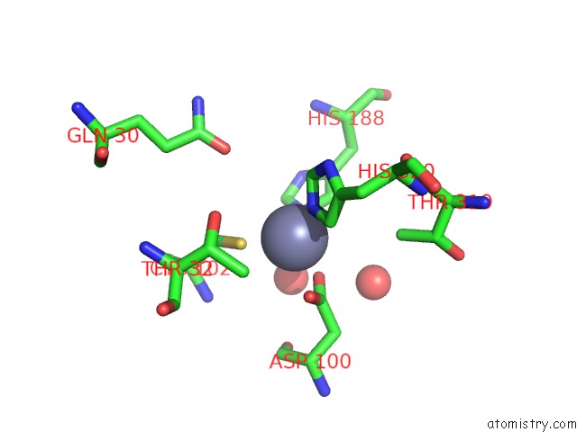
Mono view
Stereo pair view
Mono view
Stereo pair view
A full contact list of Zinc with other atoms in the Zn binding
site number 2 of Crystal Structure of A Protease-Resistant Mutant Form of Human Galectin-9 within 5.0Å range:
|
Reference:
H.Yoshida,
M.Teraoka,
N.Nishi,
S.Kamitori.
Crystal Structure of A Protease-Resistant Mutant Form of Human Galectin-9 To Be Published.
Page generated: Wed Aug 20 15:25:23 2025
Last articles
Zn in 4MM3Zn in 4MLX
Zn in 4MLT
Zn in 4MKT
Zn in 4MKP
Zn in 4MKH
Zn in 4MIM
Zn in 4MKF
Zn in 4MKG
Zn in 4MJ7