Zinc »
PDB 3kry-3l2v »
3kwc »
Zinc in PDB 3kwc: Oxidized, Active Structure of the Beta-Carboxysomal Gamma-Carbonic Anhydrase, Ccmm
Enzymatic activity of Oxidized, Active Structure of the Beta-Carboxysomal Gamma-Carbonic Anhydrase, Ccmm
All present enzymatic activity of Oxidized, Active Structure of the Beta-Carboxysomal Gamma-Carbonic Anhydrase, Ccmm:
4.2.1.1;
4.2.1.1;
Protein crystallography data
The structure of Oxidized, Active Structure of the Beta-Carboxysomal Gamma-Carbonic Anhydrase, Ccmm, PDB code: 3kwc
was solved by
M.S.Kimber,
S.E.Castel,
K.L.Pena,
with X-Ray Crystallography technique. A brief refinement statistics is given in the table below:
| Resolution Low / High (Å) | 16.88 / 2.00 |
| Space group | P 21 21 21 |
| Cell size a, b, c (Å), α, β, γ (°) | 63.900, 105.040, 196.130, 90.00, 90.00, 90.00 |
| R / Rfree (%) | 20 / 24.7 |
Other elements in 3kwc:
The structure of Oxidized, Active Structure of the Beta-Carboxysomal Gamma-Carbonic Anhydrase, Ccmm also contains other interesting chemical elements:
| Chlorine | (Cl) | 2 atoms |
Zinc Binding Sites:
The binding sites of Zinc atom in the Oxidized, Active Structure of the Beta-Carboxysomal Gamma-Carbonic Anhydrase, Ccmm
(pdb code 3kwc). This binding sites where shown within
5.0 Angstroms radius around Zinc atom.
In total 6 binding sites of Zinc where determined in the Oxidized, Active Structure of the Beta-Carboxysomal Gamma-Carbonic Anhydrase, Ccmm, PDB code: 3kwc:
Jump to Zinc binding site number: 1; 2; 3; 4; 5; 6;
In total 6 binding sites of Zinc where determined in the Oxidized, Active Structure of the Beta-Carboxysomal Gamma-Carbonic Anhydrase, Ccmm, PDB code: 3kwc:
Jump to Zinc binding site number: 1; 2; 3; 4; 5; 6;
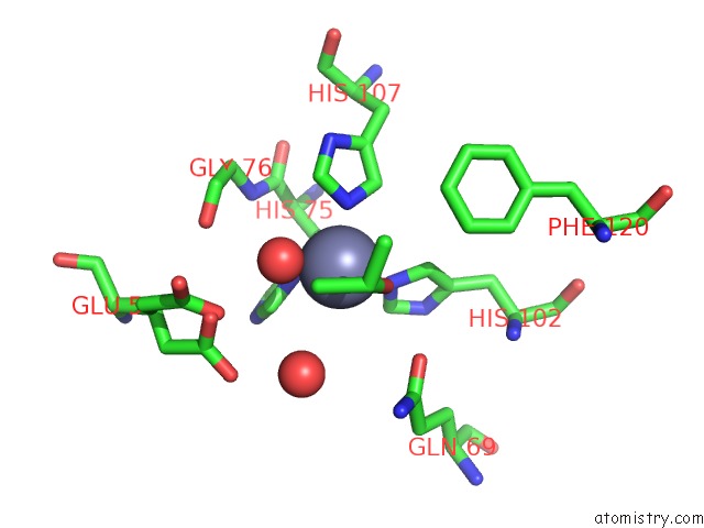
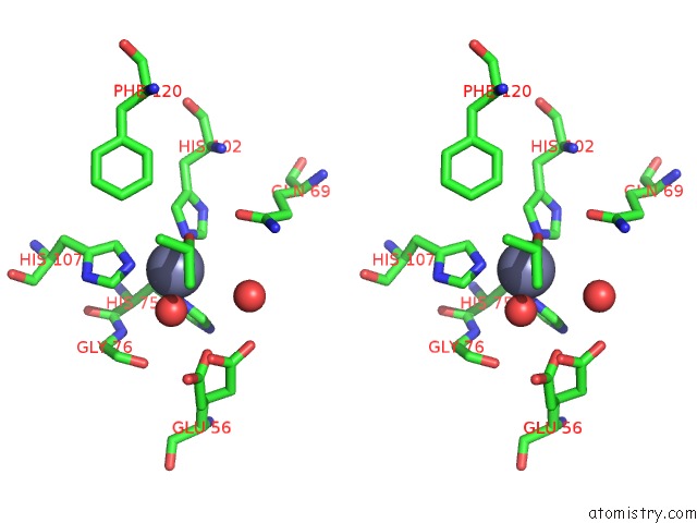
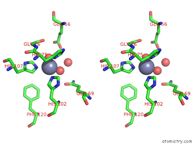
Zinc binding site 1 out of 6 in 3kwc
Go back to
Zinc binding site 1 out
of 6 in the Oxidized, Active Structure of the Beta-Carboxysomal Gamma-Carbonic Anhydrase, Ccmm
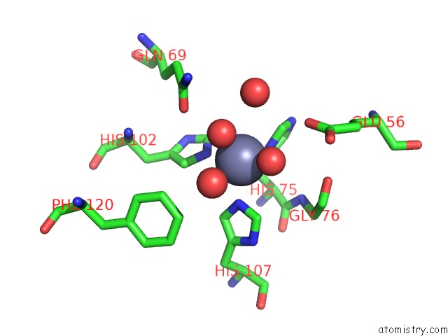
Mono view
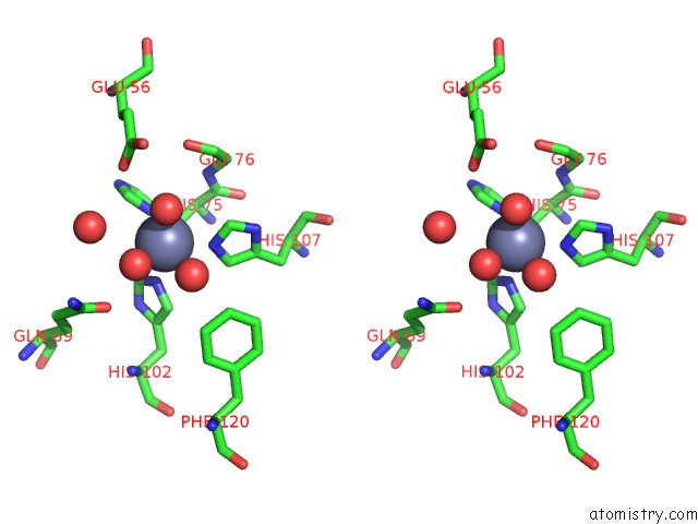
Stereo pair view
Mono view
Stereo pair view
A full contact list of Zinc with other atoms in the Zn binding
site number 1 of Oxidized, Active Structure of the Beta-Carboxysomal Gamma-Carbonic Anhydrase, Ccmm within 5.0Å range:
|
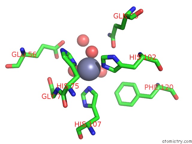
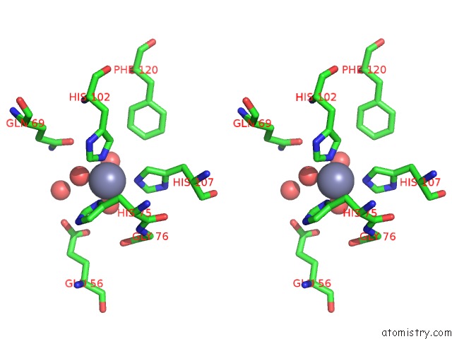
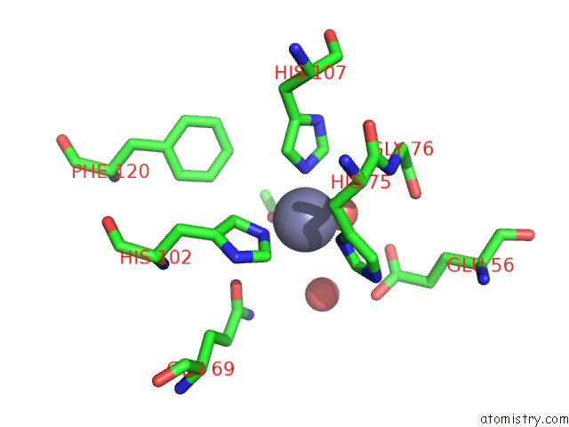
Zinc binding site 2 out of 6 in 3kwc
Go back to
Zinc binding site 2 out
of 6 in the Oxidized, Active Structure of the Beta-Carboxysomal Gamma-Carbonic Anhydrase, Ccmm
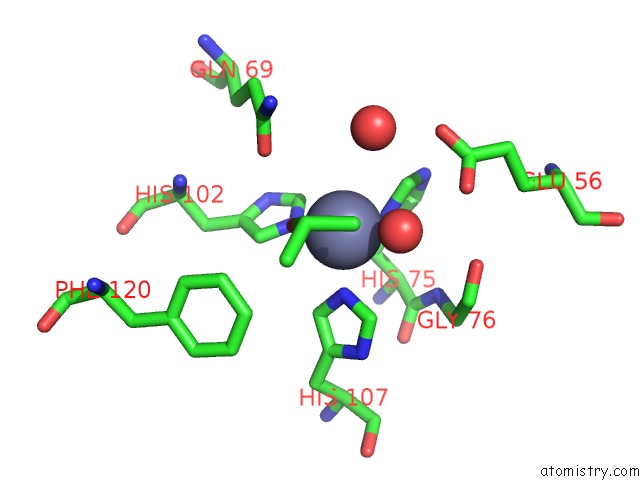
Mono view
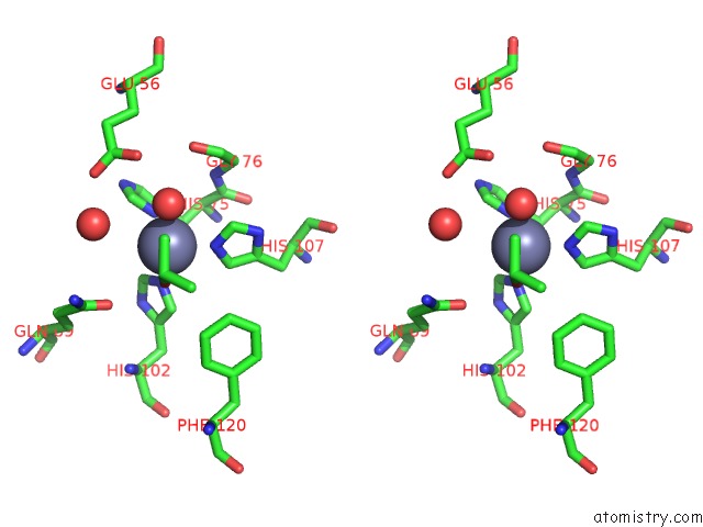
Stereo pair view
Mono view
Stereo pair view
A full contact list of Zinc with other atoms in the Zn binding
site number 2 of Oxidized, Active Structure of the Beta-Carboxysomal Gamma-Carbonic Anhydrase, Ccmm within 5.0Å range:
|
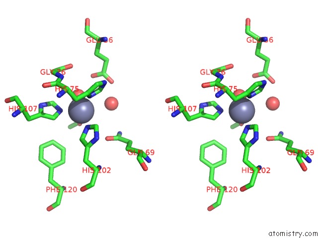
Zinc binding site 3 out of 6 in 3kwc
Go back to
Zinc binding site 3 out
of 6 in the Oxidized, Active Structure of the Beta-Carboxysomal Gamma-Carbonic Anhydrase, Ccmm
Mono view
Stereo pair view
Mono view
Stereo pair view
A full contact list of Zinc with other atoms in the Zn binding
site number 3 of Oxidized, Active Structure of the Beta-Carboxysomal Gamma-Carbonic Anhydrase, Ccmm within 5.0Å range:
|
Zinc binding site 4 out of 6 in 3kwc
Go back to
Zinc binding site 4 out
of 6 in the Oxidized, Active Structure of the Beta-Carboxysomal Gamma-Carbonic Anhydrase, Ccmm
Mono view
Stereo pair view
Mono view
Stereo pair view
A full contact list of Zinc with other atoms in the Zn binding
site number 4 of Oxidized, Active Structure of the Beta-Carboxysomal Gamma-Carbonic Anhydrase, Ccmm within 5.0Å range:
|
Zinc binding site 5 out of 6 in 3kwc
Go back to
Zinc binding site 5 out
of 6 in the Oxidized, Active Structure of the Beta-Carboxysomal Gamma-Carbonic Anhydrase, Ccmm

Mono view
Stereo pair view
Mono view
Stereo pair view
A full contact list of Zinc with other atoms in the Zn binding
site number 5 of Oxidized, Active Structure of the Beta-Carboxysomal Gamma-Carbonic Anhydrase, Ccmm within 5.0Å range:
|
Zinc binding site 6 out of 6 in 3kwc
Go back to
Zinc binding site 6 out
of 6 in the Oxidized, Active Structure of the Beta-Carboxysomal Gamma-Carbonic Anhydrase, Ccmm
Mono view
Stereo pair view
Mono view
Stereo pair view
A full contact list of Zinc with other atoms in the Zn binding
site number 6 of Oxidized, Active Structure of the Beta-Carboxysomal Gamma-Carbonic Anhydrase, Ccmm within 5.0Å range:
|
Reference:
K.L.Pena,
S.E.Castel,
C.De Araujo,
G.S.Espie,
M.S.Kimber.
Structural Basis of the Oxidative Activation of the Carboxysomal {Gamma}-Carbonic Anhydrase, Ccmm. Proc.Natl.Acad.Sci.Usa V. 107 2455 2010.
ISSN: ISSN 0027-8424
PubMed: 20133749
DOI: 10.1073/PNAS.0910866107
Page generated: Wed Aug 20 11:08:24 2025
ISSN: ISSN 0027-8424
PubMed: 20133749
DOI: 10.1073/PNAS.0910866107
Last articles
Zn in 4G9LZn in 4GAA
Zn in 4GAT
Zn in 4G9Z
Zn in 4G8H
Zn in 4G7Z
Zn in 4G7O
Zn in 4G7H
Zn in 4G83
Zn in 4G82