Atomistry » Zinc » PDB 9cbt-9fym » 9dr5
Atomistry »
  Zinc »
    PDB 9cbt-9fym »
      9dr5 »

Zinc in PDB 9dr5: Crystal Structure of Catechol 1,2-Dioxygenase From Burkholderia Multivorans (Zinc Bound, P1 Form)

Enzymatic activity of Crystal Structure of Catechol 1,2-Dioxygenase From Burkholderia Multivorans (Zinc Bound, P1 Form)

All present enzymatic activity of Crystal Structure of Catechol 1,2-Dioxygenase From Burkholderia Multivorans (Zinc Bound, P1 Form):
1.13.11.1;

Protein crystallography data

The structure of Crystal Structure of Catechol 1,2-Dioxygenase From Burkholderia Multivorans (Zinc Bound, P1 Form), PDB code: 9dr5 was solved by Seattle Structural Genomics Center For Infectious Disease, Seattlestructural Genomics Center For Infectious Disease (Ssgcid), with X-Ray Crystallography technique. A brief refinement statistics is given in the table below:

Resolution Low / High (Å) 51.99 / 1.71
Space group P 1
Cell size a, b, c (Å), α, β, γ (°) 84.97, 86.27, 110.161, 106.04, 96.74, 113.15
R / Rfree (%) 15.1 / 18.3

Zinc Binding Sites:

The binding sites of Zinc atom in the Crystal Structure of Catechol 1,2-Dioxygenase From Burkholderia Multivorans (Zinc Bound, P1 Form) (pdb code 9dr5). This binding sites where shown within 5.0 Angstroms radius around Zinc atom.
In total 8 binding sites of Zinc where determined in the Crystal Structure of Catechol 1,2-Dioxygenase From Burkholderia Multivorans (Zinc Bound, P1 Form), PDB code: 9dr5:
Jump to Zinc binding site number: 1; 2; 3; 4; 5; 6; 7; 8;

Zinc binding site 1 out of 8 in 9dr5

Go back to Zinc Binding Sites List in 9dr5
Zinc binding site 1 out of 8 in the Crystal Structure of Catechol 1,2-Dioxygenase From Burkholderia Multivorans (Zinc Bound, P1 Form)
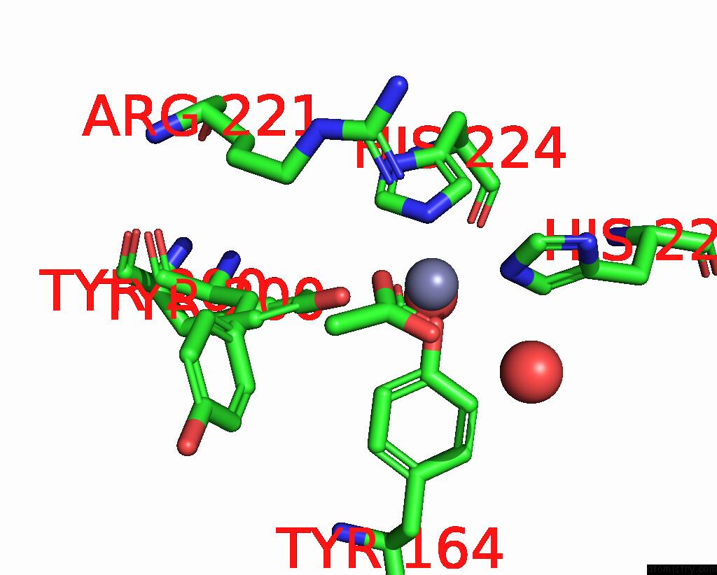

Mono view
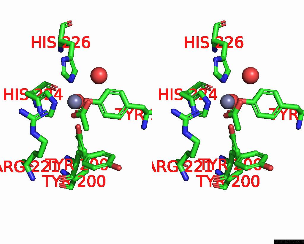

Stereo pair view

A full contact list of Zinc with other atoms in the Zn binding site number 1 of Crystal Structure of Catechol 1,2-Dioxygenase From Burkholderia Multivorans (Zinc Bound, P1 Form) within 5.0Å range:
probe atom residue distance (Å) B Occ
A:Zn401

b:27.1
occ:0.66
OXT A:ACT406 1.9 24.5 0.7
OH A:TYR164 1.9 19.5 1.0
NE2 A:HIS224 2.2 22.4 1.0
NE2 A:HIS226 2.2 21.4 1.0
OH A:TYR200 2.4 19.5 0.3
C A:ACT406 2.6 23.8 0.7
O A:ACT406 2.9 23.7 0.7
CZ A:TYR164 2.9 21.9 1.0
CD2 A:HIS226 2.9 21.9 1.0
CE1 A:HIS224 3.0 24.4 1.0
CD2 A:HIS224 3.3 27.8 1.0
CE1 A:HIS226 3.3 19.6 1.0
CE1 A:TYR164 3.6 20.3 1.0
CZ A:TYR200 3.6 18.6 0.3
NH1 A:ARG221 3.6 29.6 1.0
CE2 A:TYR164 3.8 17.9 1.0
CE2 A:TYR200 4.0 21.8 0.3
O A:HOH518 4.0 18.1 1.0
CH3 A:ACT406 4.0 22.2 0.7
CG A:HIS226 4.2 17.0 1.0
ND1 A:HIS224 4.2 24.0 1.0
ND1 A:HIS226 4.3 19.9 1.0
CG A:HIS224 4.3 20.1 1.0
O A:HOH565 4.5 19.7 1.0
CZ A:ARG221 4.7 33.2 1.0
CE1 A:TYR200 4.8 19.6 0.3
CD1 A:TYR164 4.9 22.7 1.0
CD A:ARG221 5.0 22.5 1.0

Zinc binding site 2 out of 8 in 9dr5

Go back to Zinc Binding Sites List in 9dr5
Zinc binding site 2 out of 8 in the Crystal Structure of Catechol 1,2-Dioxygenase From Burkholderia Multivorans (Zinc Bound, P1 Form)
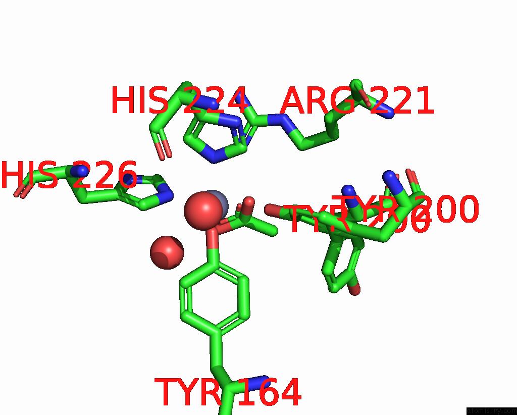

Mono view
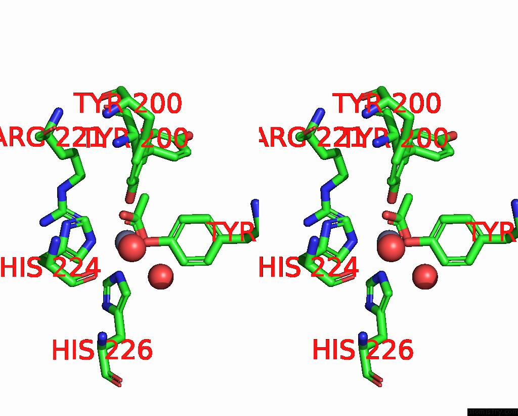

Stereo pair view

A full contact list of Zinc with other atoms in the Zn binding site number 2 of Crystal Structure of Catechol 1,2-Dioxygenase From Burkholderia Multivorans (Zinc Bound, P1 Form) within 5.0Å range:
probe atom residue distance (Å) B Occ
B:Zn401

b:23.3
occ:0.52
OH B:TYR164 1.8 18.9 1.0
OXT B:ACT404 2.0 24.7 0.5
NE2 B:HIS226 2.2 22.7 1.0
NE2 B:HIS224 2.3 20.2 1.0
OH B:TYR200 2.5 18.6 0.5
C B:ACT404 2.6 21.8 0.5
O B:ACT404 2.7 17.8 0.5
CZ B:TYR164 2.9 18.8 1.0
CD2 B:HIS226 2.9 21.6 1.0
CE1 B:HIS224 3.1 22.3 1.0
CE1 B:HIS226 3.3 25.0 1.0
CD2 B:HIS224 3.3 25.8 1.0
CE1 B:TYR164 3.7 20.7 1.0
CZ B:TYR200 3.7 20.9 0.5
CE2 B:TYR164 3.7 17.0 1.0
NH1 B:ARG221 3.7 29.0 1.0
O B:HOH538 4.0 16.8 1.0
CH3 B:ACT404 4.0 22.1 0.5
CE2 B:TYR200 4.2 20.6 0.5
CG B:HIS226 4.2 17.9 1.0
ND1 B:HIS224 4.2 21.9 1.0
ND1 B:HIS226 4.3 20.4 1.0
CG B:HIS224 4.4 20.0 1.0
O B:HOH627 4.5 17.1 1.0
CZ B:ARG221 4.8 27.2 1.0
CE1 B:TYR200 4.8 21.0 0.5
CD1 B:TYR164 4.9 20.5 1.0
CD2 B:TYR164 5.0 17.6 1.0

Zinc binding site 3 out of 8 in 9dr5

Go back to Zinc Binding Sites List in 9dr5
Zinc binding site 3 out of 8 in the Crystal Structure of Catechol 1,2-Dioxygenase From Burkholderia Multivorans (Zinc Bound, P1 Form)
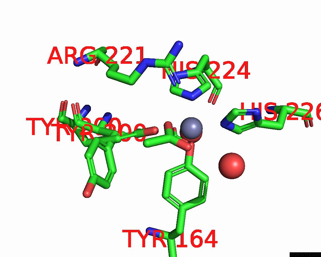

Mono view
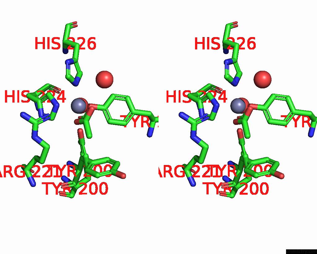

Stereo pair view

A full contact list of Zinc with other atoms in the Zn binding site number 3 of Crystal Structure of Catechol 1,2-Dioxygenase From Burkholderia Multivorans (Zinc Bound, P1 Form) within 5.0Å range:
probe atom residue distance (Å) B Occ
C:Zn401

b:29.7
occ:0.69
OH C:TYR164 1.9 22.4 1.0
OXT C:ACT406 2.0 22.9 0.7
NE2 C:HIS226 2.2 22.1 1.0
NE2 C:HIS224 2.3 23.9 1.0
OH C:TYR200 2.3 21.6 0.3
C C:ACT406 2.7 22.8 0.7
O C:ACT406 2.9 22.1 0.7
CZ C:TYR164 2.9 21.6 1.0
CD2 C:HIS226 3.0 21.7 1.0
CE1 C:HIS224 3.0 28.5 1.0
CE1 C:HIS226 3.3 21.0 1.0
CD2 C:HIS224 3.4 28.5 1.0
CZ C:TYR200 3.6 22.5 0.3
CE1 C:TYR164 3.6 22.4 1.0
NH1 C:ARG221 3.7 29.2 1.0
CE2 C:TYR164 3.8 19.9 1.0
O C:HOH519 4.0 20.0 1.0
CE2 C:TYR200 4.0 22.5 0.3
CH3 C:ACT406 4.1 21.2 0.7
CG C:HIS226 4.2 22.6 1.0
ND1 C:HIS224 4.2 25.8 1.0
ND1 C:HIS226 4.3 22.1 1.0
CG C:HIS224 4.4 20.8 1.0
O C:HOH648 4.5 19.8 1.0
CE1 C:TYR200 4.7 19.6 0.3
CZ C:ARG221 4.7 26.1 1.0
CD1 C:TYR164 4.9 22.5 1.0
CD C:ARG221 5.0 26.5 1.0

Zinc binding site 4 out of 8 in 9dr5

Go back to Zinc Binding Sites List in 9dr5
Zinc binding site 4 out of 8 in the Crystal Structure of Catechol 1,2-Dioxygenase From Burkholderia Multivorans (Zinc Bound, P1 Form)
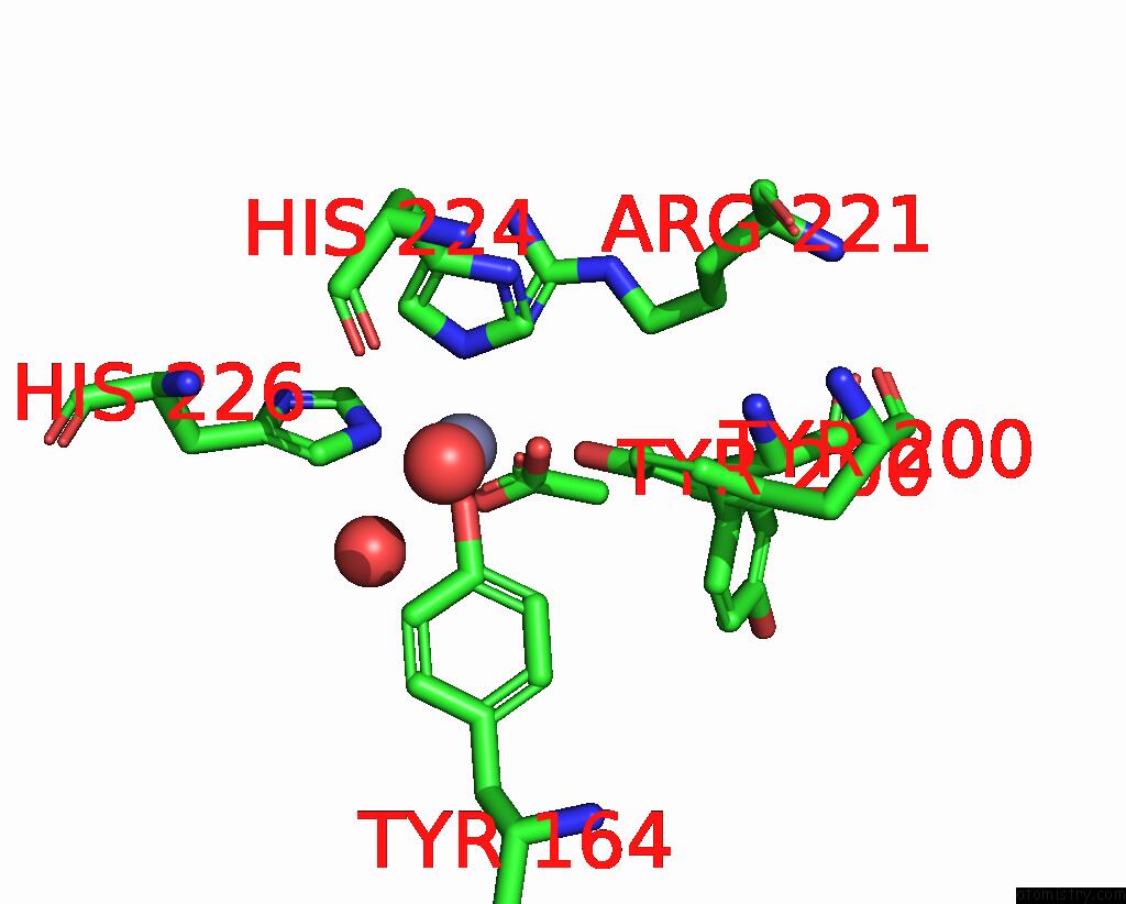

Mono view
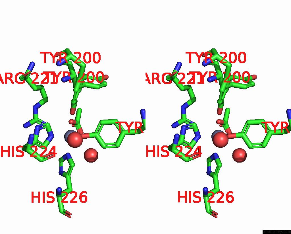

Stereo pair view

A full contact list of Zinc with other atoms in the Zn binding site number 4 of Crystal Structure of Catechol 1,2-Dioxygenase From Burkholderia Multivorans (Zinc Bound, P1 Form) within 5.0Å range:
probe atom residue distance (Å) B Occ
D:Zn401

b:23.4
occ:0.57
OH D:TYR164 1.9 17.1 1.0
OXT D:ACT404 1.9 21.7 0.6
NE2 D:HIS226 2.1 19.0 1.0
NE2 D:HIS224 2.2 21.5 1.0
OH D:TYR200 2.5 15.2 0.4
C D:ACT404 2.7 24.4 0.6
CD2 D:HIS226 2.9 20.6 1.0
O D:ACT404 2.9 18.1 0.6
CZ D:TYR164 2.9 16.0 1.0
CE1 D:HIS224 3.1 21.7 1.0
CE1 D:HIS226 3.3 20.3 1.0
CD2 D:HIS224 3.3 21.6 1.0
NH1 D:ARG221 3.6 29.5 1.0
CE1 D:TYR164 3.7 17.2 1.0
CE2 D:TYR164 3.8 14.6 1.0
CZ D:TYR200 3.8 14.1 0.4
O D:HOH543 4.0 17.3 1.0
CG D:HIS226 4.1 15.4 1.0
CH3 D:ACT404 4.1 15.9 0.6
ND1 D:HIS224 4.2 20.4 1.0
ND1 D:HIS226 4.2 18.5 1.0
CE2 D:TYR200 4.3 16.6 0.4
CG D:HIS224 4.3 17.9 1.0
O D:HOH627 4.5 15.5 1.0
CZ D:ARG221 4.7 23.8 1.0
CE1 D:TYR200 4.9 17.6 0.4
CD D:ARG221 5.0 20.0 1.0
CD1 D:TYR164 5.0 17.0 1.0

Zinc binding site 5 out of 8 in 9dr5

Go back to Zinc Binding Sites List in 9dr5
Zinc binding site 5 out of 8 in the Crystal Structure of Catechol 1,2-Dioxygenase From Burkholderia Multivorans (Zinc Bound, P1 Form)
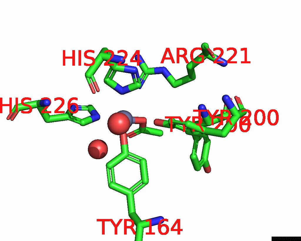

Mono view
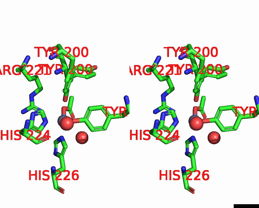

Stereo pair view

A full contact list of Zinc with other atoms in the Zn binding site number 5 of Crystal Structure of Catechol 1,2-Dioxygenase From Burkholderia Multivorans (Zinc Bound, P1 Form) within 5.0Å range:
probe atom residue distance (Å) B Occ
E:Zn401

b:24.7
occ:0.67
OXT E:ACT404 1.8 23.7 0.7
OH E:TYR164 1.8 18.4 1.0
NE2 E:HIS226 2.2 17.9 1.0
NE2 E:HIS224 2.3 23.6 1.0
OH E:TYR200 2.5 20.3 0.3
C E:ACT404 2.6 23.6 0.7
O E:ACT404 2.9 19.9 0.7
CZ E:TYR164 2.9 17.1 1.0
CD2 E:HIS226 3.0 18.0 1.0
CE1 E:HIS224 3.1 27.2 1.0
CE1 E:HIS226 3.3 18.8 1.0
CD2 E:HIS224 3.3 21.8 1.0
CE1 E:TYR164 3.6 16.1 1.0
NH1 E:ARG221 3.6 23.6 1.0
CZ E:TYR200 3.7 18.0 0.3
CE2 E:TYR164 3.8 17.1 1.0
O E:HOH520 3.9 16.2 1.0
CH3 E:ACT404 4.0 17.6 0.7
CE2 E:TYR200 4.1 20.6 0.3
CG E:HIS226 4.2 14.9 1.0
ND1 E:HIS224 4.2 20.8 1.0
ND1 E:HIS226 4.3 15.3 1.0
CG E:HIS224 4.4 17.2 1.0
O E:HOH612 4.5 14.3 1.0
CZ E:ARG221 4.7 24.5 1.0
CE1 E:TYR200 4.9 19.1 0.3
CD1 E:TYR164 4.9 17.0 1.0
CD E:ARG221 5.0 22.5 1.0

Zinc binding site 6 out of 8 in 9dr5

Go back to Zinc Binding Sites List in 9dr5
Zinc binding site 6 out of 8 in the Crystal Structure of Catechol 1,2-Dioxygenase From Burkholderia Multivorans (Zinc Bound, P1 Form)
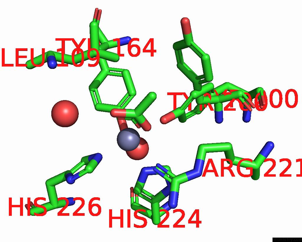

Mono view
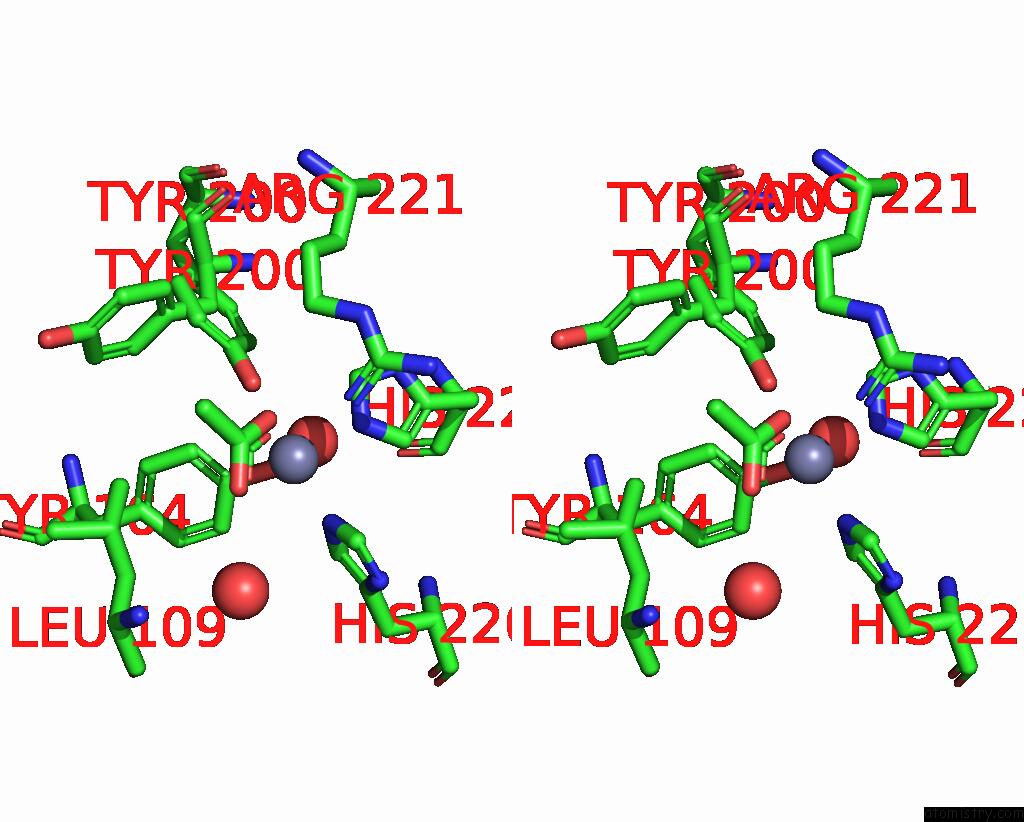

Stereo pair view

A full contact list of Zinc with other atoms in the Zn binding site number 6 of Crystal Structure of Catechol 1,2-Dioxygenase From Burkholderia Multivorans (Zinc Bound, P1 Form) within 5.0Å range:
probe atom residue distance (Å) B Occ
F:Zn401

b:28.4
occ:0.64
OH F:TYR164 1.8 21.8 1.0
OXT F:ACT406 1.8 26.9 0.6
NE2 F:HIS226 2.2 19.3 1.0
NE2 F:HIS224 2.2 23.2 1.0
OH F:TYR200 2.5 20.3 0.4
C F:ACT406 2.6 28.1 0.6
O F:ACT406 2.8 22.3 0.6
CZ F:TYR164 2.9 20.0 1.0
CD2 F:HIS226 3.0 23.2 1.0
CE1 F:HIS224 3.0 30.7 1.0
CE1 F:HIS226 3.3 22.9 1.0
CD2 F:HIS224 3.3 27.9 1.0
CE1 F:TYR164 3.5 21.3 1.0
NH1 F:ARG221 3.6 33.1 1.0
CZ F:TYR200 3.7 22.9 0.4
CE2 F:TYR164 3.8 18.4 1.0
CH3 F:ACT406 4.0 24.1 0.6
O F:HOH565 4.0 21.4 1.0
CE2 F:TYR200 4.1 19.9 0.4
CG F:HIS226 4.2 18.6 1.0
ND1 F:HIS224 4.2 24.8 1.0
ND1 F:HIS226 4.3 21.5 1.0
CG F:HIS224 4.4 24.4 1.0
O F:HOH615 4.6 17.8 1.0
CZ F:ARG221 4.7 33.6 1.0
CE1 F:TYR200 4.8 22.7 0.4
CD1 F:TYR164 4.8 22.5 1.0
CD F:ARG221 5.0 23.2 1.0
CD1 F:LEU109 5.0 28.9 1.0

Zinc binding site 7 out of 8 in 9dr5

Go back to Zinc Binding Sites List in 9dr5
Zinc binding site 7 out of 8 in the Crystal Structure of Catechol 1,2-Dioxygenase From Burkholderia Multivorans (Zinc Bound, P1 Form)
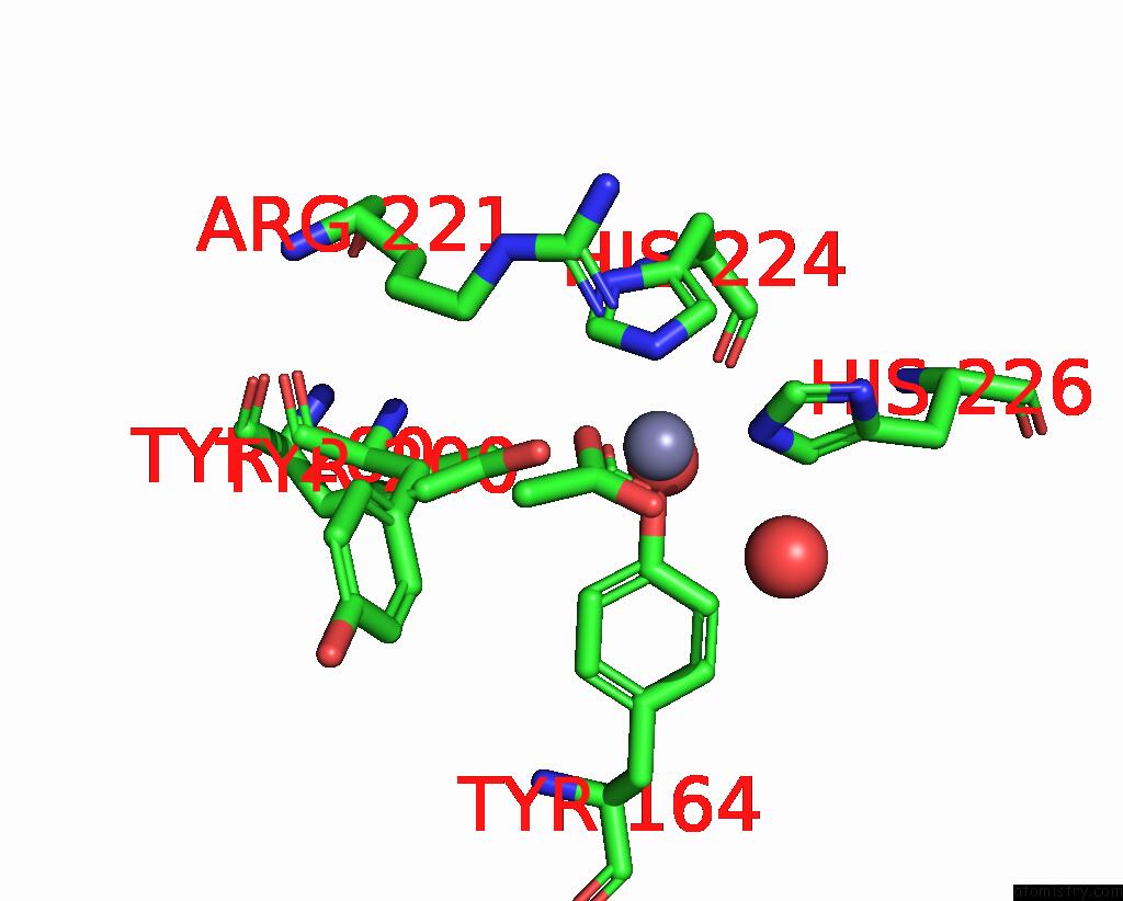

Mono view
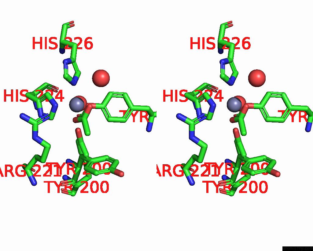

Stereo pair view

A full contact list of Zinc with other atoms in the Zn binding site number 7 of Crystal Structure of Catechol 1,2-Dioxygenase From Burkholderia Multivorans (Zinc Bound, P1 Form) within 5.0Å range:
probe atom residue distance (Å) B Occ
G:Zn401

b:23.1
occ:0.55
OH G:TYR164 1.8 17.5 1.0
OXT G:ACT405 1.9 23.2 0.6
NE2 G:HIS226 2.2 20.6 1.0
NE2 G:HIS224 2.2 18.4 1.0
OH G:TYR200 2.4 19.7 0.5
C G:ACT405 2.6 20.6 0.6
O G:ACT405 2.8 16.0 0.6
CZ G:TYR164 2.9 20.0 1.0
CD2 G:HIS226 3.0 18.9 1.0
CE1 G:HIS224 3.1 22.9 1.0
CE1 G:HIS226 3.2 23.2 1.0
CD2 G:HIS224 3.3 21.7 1.0
NH1 G:ARG221 3.7 27.7 1.0
CZ G:TYR200 3.7 18.3 0.5
CE1 G:TYR164 3.7 16.2 1.0
CE2 G:TYR164 3.8 18.0 1.0
O G:HOH576 4.0 17.7 1.0
CH3 G:ACT405 4.1 18.9 0.6
CE1 G:TYR200 4.2 14.3 0.5
CG G:HIS226 4.2 14.9 1.0
ND1 G:HIS224 4.2 18.4 1.0
ND1 G:HIS226 4.3 19.0 1.0
CG G:HIS224 4.3 16.9 1.0
O G:HOH628 4.6 16.3 1.0
CZ G:ARG221 4.7 22.3 1.0
CE2 G:TYR200 4.8 20.2 0.5
CD G:ARG221 4.9 18.8 1.0
CD1 G:TYR164 5.0 20.9 1.0

Zinc binding site 8 out of 8 in 9dr5

Go back to Zinc Binding Sites List in 9dr5
Zinc binding site 8 out of 8 in the Crystal Structure of Catechol 1,2-Dioxygenase From Burkholderia Multivorans (Zinc Bound, P1 Form)
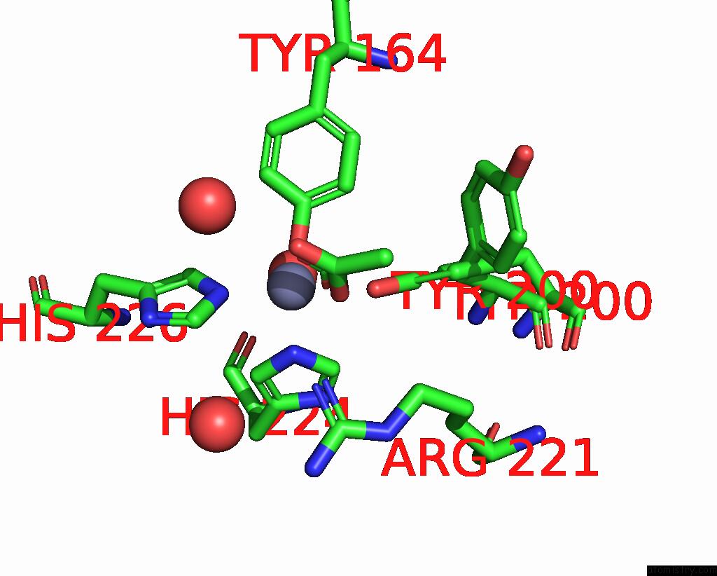

Mono view
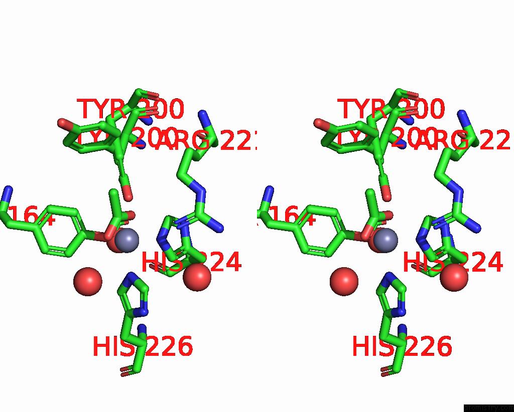

Stereo pair view

A full contact list of Zinc with other atoms in the Zn binding site number 8 of Crystal Structure of Catechol 1,2-Dioxygenase From Burkholderia Multivorans (Zinc Bound, P1 Form) within 5.0Å range:
probe atom residue distance (Å) B Occ
H:Zn401

b:24.6
occ:0.59
OXT H:ACT405 1.8 24.3 0.6
OH H:TYR164 1.9 19.9 1.0
NE2 H:HIS226 2.2 19.4 1.0
NE2 H:HIS224 2.2 20.8 1.0
OH H:TYR200 2.5 14.7 0.4
C H:ACT405 2.6 24.9 0.6
O H:ACT405 2.8 20.5 0.6
CZ H:TYR164 2.9 17.5 1.0
CD2 H:HIS226 2.9 22.6 1.0
CE1 H:HIS224 3.1 26.5 1.0
CD2 H:HIS224 3.3 21.3 1.0
CE1 H:HIS226 3.3 23.5 1.0
CE1 H:TYR164 3.7 18.0 1.0
NH1 H:ARG221 3.7 22.9 1.0
CZ H:TYR200 3.7 17.0 0.4
CE2 H:TYR164 3.8 17.8 1.0
O H:HOH532 4.0 17.7 1.0
CH3 H:ACT405 4.0 17.9 0.6
CE2 H:TYR200 4.1 15.2 0.4
CG H:HIS226 4.2 17.3 1.0
ND1 H:HIS224 4.3 19.7 1.0
ND1 H:HIS226 4.3 20.3 1.0
CG H:HIS224 4.4 16.7 1.0
O H:HOH566 4.6 15.6 1.0
CZ H:ARG221 4.7 21.1 1.0
CE1 H:TYR200 4.8 19.4 0.4
CD1 H:TYR164 4.9 18.2 1.0
O H:HOH501 5.0 26.6 1.0

Reference:

P.Enayati, L.Liu, S.Lovell, G.W.Buchko, K.P.Battaile. Crystal Structure of Catechol 1,2-Dioxygenase From Burkholderia Multivorans (Zinc Bound, P1 Form) To Be Published.
Page generated: Thu Oct 31 15:26:07 2024

Last articles

W in 8QLN
W in 8RJA
V in 8WTN
Te in 8QLN
Re in 9GHX
Rb in 8Z5C
Ni in 9C0T
Ni in 9C0S
Ni in 9GP1
Ni in 9FYO
© Copyright 2008-2020 by atomistry.com
Home   |    Site Map   |    Copyright   |    Contact us   |    Privacy