Zinc in PDB 9d60: Crystal Structure of Zn(II)-Bound Polysaccharide Deacetylase From Bacteroides Ovatus
Protein crystallography data
The structure of Crystal Structure of Zn(II)-Bound Polysaccharide Deacetylase From Bacteroides Ovatus, PDB code: 9d60
was solved by
O.E.Adamek,
K.J.Mclaughlin,
with X-Ray Crystallography technique. A brief refinement statistics is given in the table below:
| Resolution Low / High (Å) | 47.69 / 1.36 |
| Space group | P 21 21 21 |
| Cell size a, b, c (Å), α, β, γ (°) | 69.295, 73.112, 95.385, 90, 90, 90 |
| R / Rfree (%) | 16.5 / 17.7 |
Zinc Binding Sites:
The binding sites of Zinc atom in the Crystal Structure of Zn(II)-Bound Polysaccharide Deacetylase From Bacteroides Ovatus
(pdb code 9d60). This binding sites where shown within
5.0 Angstroms radius around Zinc atom.
In total only one binding site of Zinc was determined in the Crystal Structure of Zn(II)-Bound Polysaccharide Deacetylase From Bacteroides Ovatus, PDB code: 9d60:
In total only one binding site of Zinc was determined in the Crystal Structure of Zn(II)-Bound Polysaccharide Deacetylase From Bacteroides Ovatus, PDB code: 9d60:
Zinc binding site 1 out of 1 in 9d60
Go back to
Zinc binding site 1 out
of 1 in the Crystal Structure of Zn(II)-Bound Polysaccharide Deacetylase From Bacteroides Ovatus
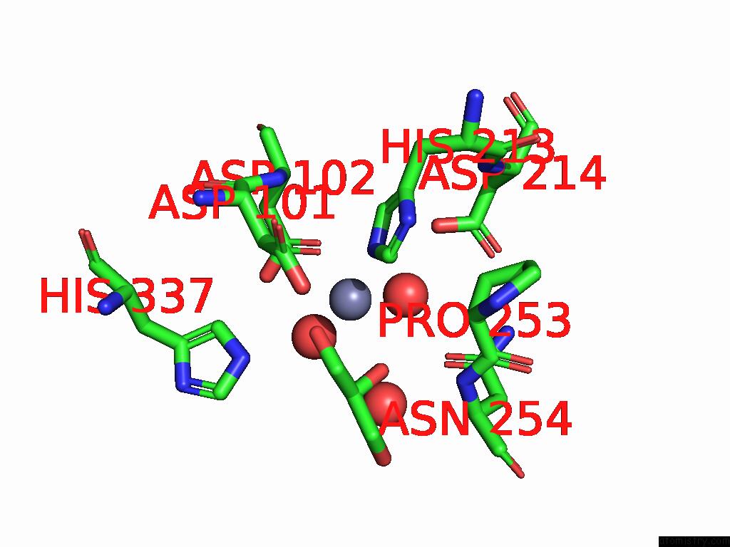
Mono view
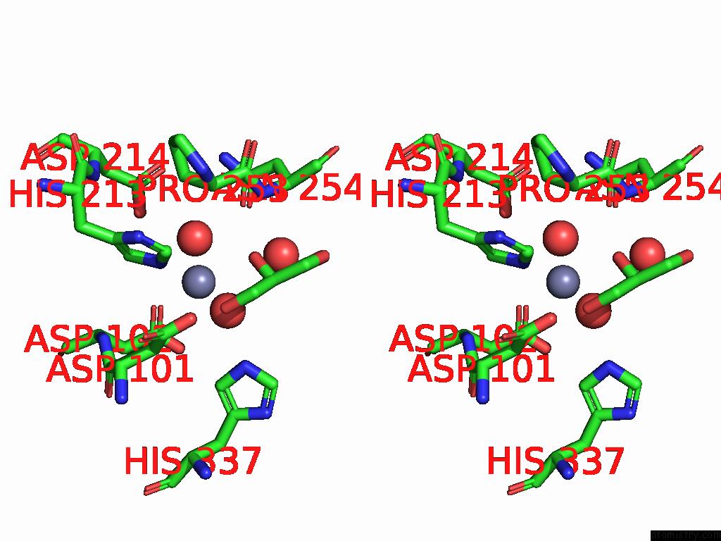
Stereo pair view
Mono view
Stereo pair view
A full contact list of Zinc with other atoms in the Zn binding
site number 1 of Crystal Structure of Zn(II)-Bound Polysaccharide Deacetylase From Bacteroides Ovatus within 5.0Å range:
|
Reference:
L.A.Schwartz,
J.O.Norman,
S.Hasan,
O.E.Adamek,
E.Dzuong,
J.C.Lowenstein,
O.G.Yost,
B.Sankaran,
K.J.Mclaughlin.
Carbohydrate Deacetylase Unique to Gut Microbe Bacteroides Reveals Atypical Structure. Biochemistry 2024.
ISSN: ISSN 0006-2960
PubMed: 39663570
DOI: 10.1021/ACS.BIOCHEM.4C00519
Page generated: Sun Feb 9 01:02:05 2025
ISSN: ISSN 0006-2960
PubMed: 39663570
DOI: 10.1021/ACS.BIOCHEM.4C00519
Last articles
Zn in 9MJ5Zn in 9HNW
Zn in 9G0L
Zn in 9FNE
Zn in 9DZN
Zn in 9E0I
Zn in 9D32
Zn in 9DAK
Zn in 8ZXC
Zn in 8ZUF