Zinc in PDB 9ckg: Crystal Structure of SMYD2 Active Site Mutant
Enzymatic activity of Crystal Structure of SMYD2 Active Site Mutant
All present enzymatic activity of Crystal Structure of SMYD2 Active Site Mutant:
2.1.1.354;
2.1.1.354;
Protein crystallography data
The structure of Crystal Structure of SMYD2 Active Site Mutant, PDB code: 9ckg
was solved by
J.Sobota,
Y.Zhang,
N.Spellmon,
E.Perry,
J.Brunzelle,
Z.Yang,
with X-Ray Crystallography technique. A brief refinement statistics is given in the table below:
| Resolution Low / High (Å) | 48.01 / 2.75 |
| Space group | I 4 |
| Cell size a, b, c (Å), α, β, γ (°) | 151.822, 151.822, 53.377, 90, 90, 90 |
| R / Rfree (%) | 19.4 / 22.3 |
Zinc Binding Sites:
The binding sites of Zinc atom in the Crystal Structure of SMYD2 Active Site Mutant
(pdb code 9ckg). This binding sites where shown within
5.0 Angstroms radius around Zinc atom.
In total 3 binding sites of Zinc where determined in the Crystal Structure of SMYD2 Active Site Mutant, PDB code: 9ckg:
Jump to Zinc binding site number: 1; 2; 3;
In total 3 binding sites of Zinc where determined in the Crystal Structure of SMYD2 Active Site Mutant, PDB code: 9ckg:
Jump to Zinc binding site number: 1; 2; 3;
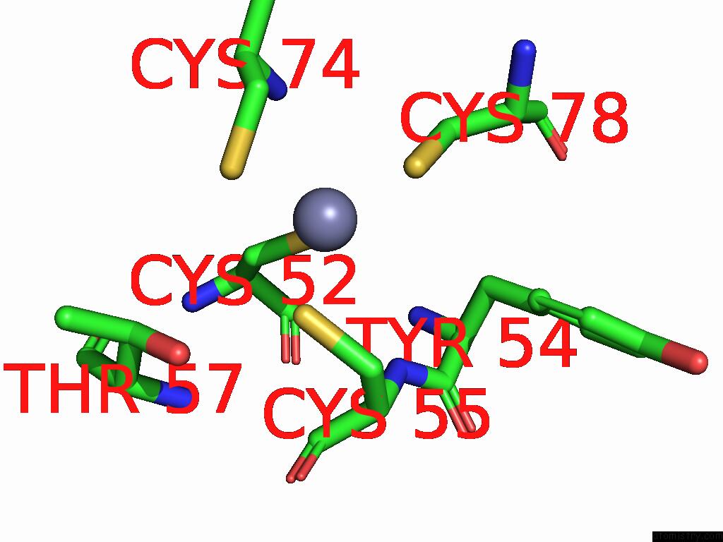
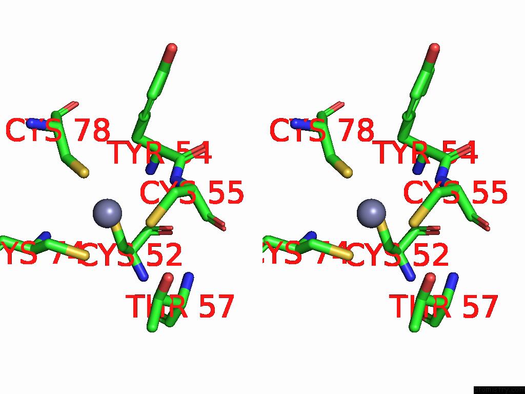
Zinc binding site 1 out of 3 in 9ckg
Go back to
Zinc binding site 1 out
of 3 in the Crystal Structure of SMYD2 Active Site Mutant
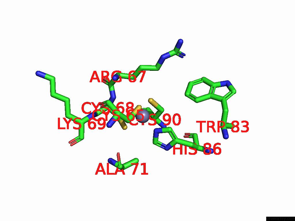
Mono view
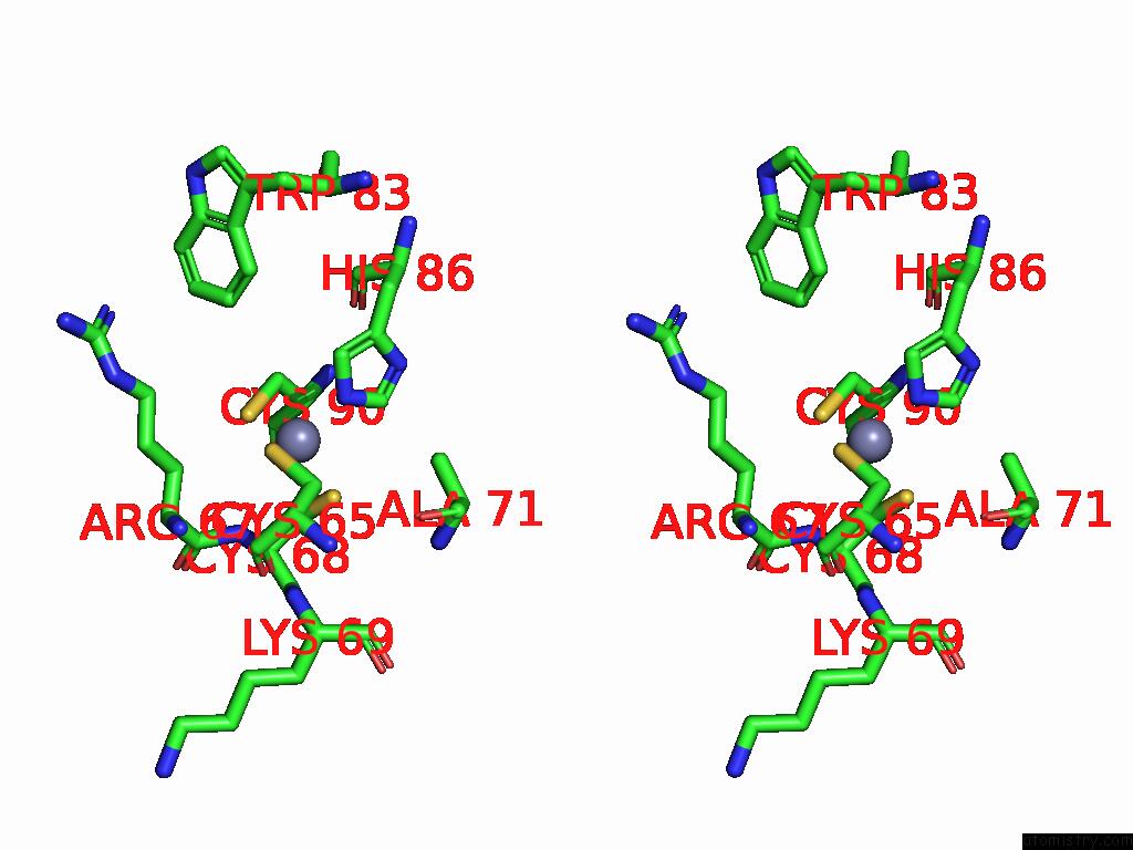
Stereo pair view
Mono view
Stereo pair view
A full contact list of Zinc with other atoms in the Zn binding
site number 1 of Crystal Structure of SMYD2 Active Site Mutant within 5.0Å range:
|
Zinc binding site 2 out of 3 in 9ckg
Go back to
Zinc binding site 2 out
of 3 in the Crystal Structure of SMYD2 Active Site Mutant
Mono view
Stereo pair view
Mono view
Stereo pair view
A full contact list of Zinc with other atoms in the Zn binding
site number 2 of Crystal Structure of SMYD2 Active Site Mutant within 5.0Å range:
|
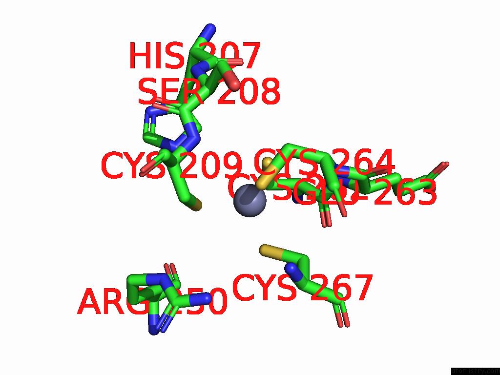
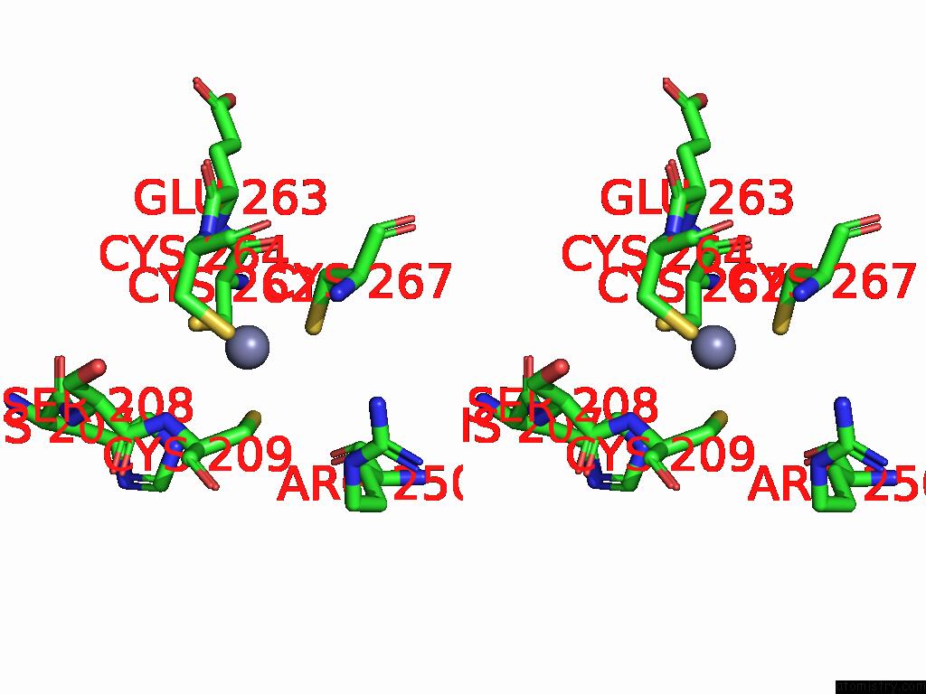
Zinc binding site 3 out of 3 in 9ckg
Go back to
Zinc binding site 3 out
of 3 in the Crystal Structure of SMYD2 Active Site Mutant
Mono view
Stereo pair view
Mono view
Stereo pair view
A full contact list of Zinc with other atoms in the Zn binding
site number 3 of Crystal Structure of SMYD2 Active Site Mutant within 5.0Å range:
|
Reference:
Y.Zhang,
E.Alshammari,
J.Sobota,
N.Spellmon,
E.Perry,
T.Cao,
T.Mugunamalwaththa,
S.Smith,
J.Brunzelle,
G.Wu,
T.Stemmler,
J.Jin,
C.Li,
Z.Yang.
Structure of the SMYD2-PARP1 Complex Reveals Both Productive and Allosteric Modes of Peptide Binding. Biorxiv 2024.
ISSN: ISSN 2692-8205
PubMed: 39677743
DOI: 10.1101/2024.12.03.626679
Page generated: Sun Feb 9 01:02:05 2025
ISSN: ISSN 2692-8205
PubMed: 39677743
DOI: 10.1101/2024.12.03.626679
Last articles
Zn in 9MJ5Zn in 9HNW
Zn in 9G0L
Zn in 9FNE
Zn in 9DZN
Zn in 9E0I
Zn in 9D32
Zn in 9DAK
Zn in 8ZXC
Zn in 8ZUF