Zinc in PDB 9azj: Structure of Ubiquitinated Nemo Uban K285C-Ub G76C Bound to Hoip NZF1
Enzymatic activity of Structure of Ubiquitinated Nemo Uban K285C-Ub G76C Bound to Hoip NZF1
All present enzymatic activity of Structure of Ubiquitinated Nemo Uban K285C-Ub G76C Bound to Hoip NZF1:
2.3.2.31;
2.3.2.31;
Protein crystallography data
The structure of Structure of Ubiquitinated Nemo Uban K285C-Ub G76C Bound to Hoip NZF1, PDB code: 9azj
was solved by
M.A.Michel,
S.Scutts,
D.Komander,
with X-Ray Crystallography technique. A brief refinement statistics is given in the table below:
| Resolution Low / High (Å) | 50.72 / 3.32 |
| Space group | P 21 21 21 |
| Cell size a, b, c (Å), α, β, γ (°) | 66.828, 101.447, 144.14, 90, 90, 90 |
| R / Rfree (%) | 24.1 / 28.5 |
Zinc Binding Sites:
The binding sites of Zinc atom in the Structure of Ubiquitinated Nemo Uban K285C-Ub G76C Bound to Hoip NZF1
(pdb code 9azj). This binding sites where shown within
5.0 Angstroms radius around Zinc atom.
In total 2 binding sites of Zinc where determined in the Structure of Ubiquitinated Nemo Uban K285C-Ub G76C Bound to Hoip NZF1, PDB code: 9azj:
Jump to Zinc binding site number: 1; 2;
In total 2 binding sites of Zinc where determined in the Structure of Ubiquitinated Nemo Uban K285C-Ub G76C Bound to Hoip NZF1, PDB code: 9azj:
Jump to Zinc binding site number: 1; 2;
Zinc binding site 1 out of 2 in 9azj
Go back to
Zinc binding site 1 out
of 2 in the Structure of Ubiquitinated Nemo Uban K285C-Ub G76C Bound to Hoip NZF1
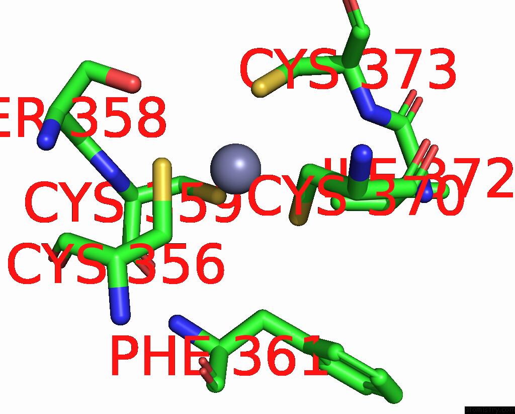
Mono view
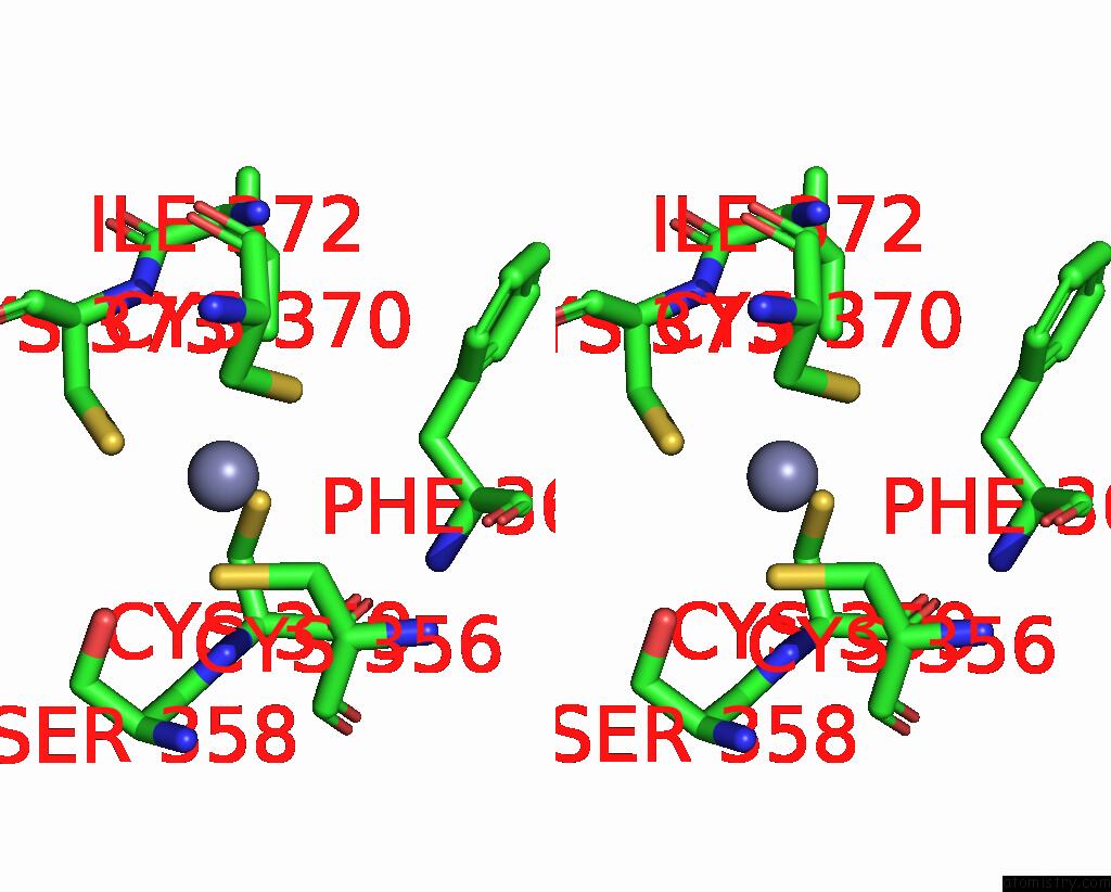
Stereo pair view
Mono view
Stereo pair view
A full contact list of Zinc with other atoms in the Zn binding
site number 1 of Structure of Ubiquitinated Nemo Uban K285C-Ub G76C Bound to Hoip NZF1 within 5.0Å range:
|
Zinc binding site 2 out of 2 in 9azj
Go back to
Zinc binding site 2 out
of 2 in the Structure of Ubiquitinated Nemo Uban K285C-Ub G76C Bound to Hoip NZF1
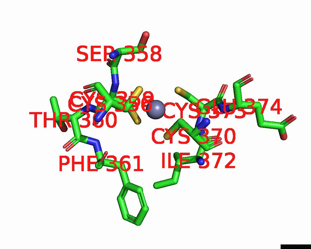
Mono view
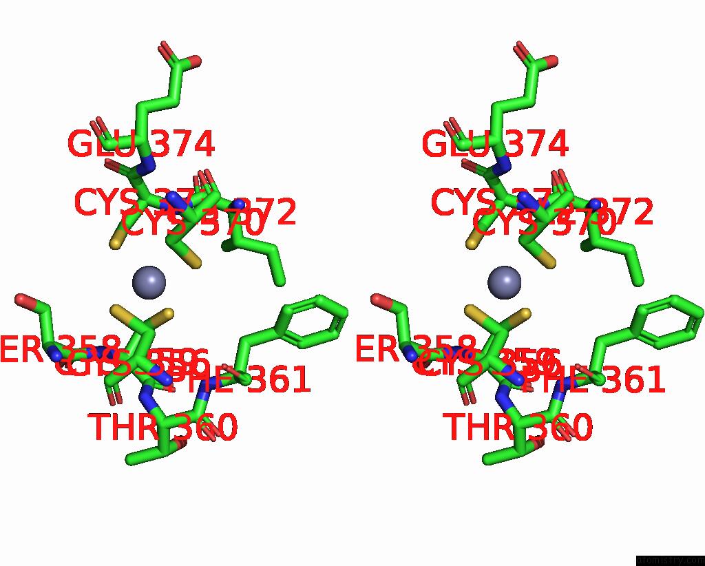
Stereo pair view
Mono view
Stereo pair view
A full contact list of Zinc with other atoms in the Zn binding
site number 2 of Structure of Ubiquitinated Nemo Uban K285C-Ub G76C Bound to Hoip NZF1 within 5.0Å range:
|
Reference:
M.A.Michel,
S.Scutts,
D.Komander.
Structure of TAB2 Nzf Domain Bound to K6 / LYS6-Linked Diubiquitin To Be Published.
Page generated: Thu Oct 31 14:37:32 2024
Last articles
Zn in 9MJ5Zn in 9HNW
Zn in 9G0L
Zn in 9FNE
Zn in 9DZN
Zn in 9E0I
Zn in 9D32
Zn in 9DAK
Zn in 8ZXC
Zn in 8ZUF