Zinc in PDB 8zu5: Crystal Structure of Methyl Parathion Hydrolase Mutant A66D/I143V/I145L/Q272D/S279A/
Protein crystallography data
The structure of Crystal Structure of Methyl Parathion Hydrolase Mutant A66D/I143V/I145L/Q272D/S279A/, PDB code: 8zu5
was solved by
F.Xu,
S.Fan,
with X-Ray Crystallography technique. A brief refinement statistics is given in the table below:
Resolution Low / High (Å)
|
29.32 /
1.83
|
Space group
|
C 1 2 1
|
Cell size a, b, c (Å), α, β, γ (°)
|
58.801,
83.719,
115.492,
90,
94.33,
90
|
R / Rfree (%)
|
20.8 /
24.2
|
Zinc Binding Sites:
The binding sites of Zinc atom in the Crystal Structure of Methyl Parathion Hydrolase Mutant A66D/I143V/I145L/Q272D/S279A/
(pdb code 8zu5). This binding sites where shown within
5.0 Angstroms radius around Zinc atom.
In total 4 binding sites of Zinc where determined in the
Crystal Structure of Methyl Parathion Hydrolase Mutant A66D/I143V/I145L/Q272D/S279A/, PDB code: 8zu5:
Jump to Zinc binding site number:
1;
2;
3;
4;
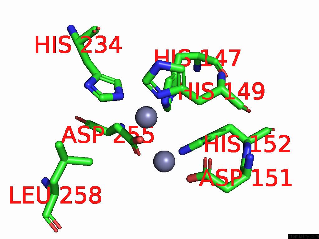
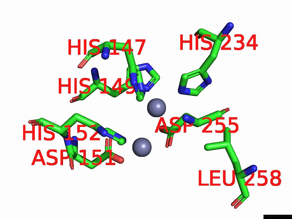
Zinc binding site 1 out
of 4 in 8zu5
Go back to
Zinc Binding Sites List in 8zu5
Zinc binding site 1 out
of 4 in the Crystal Structure of Methyl Parathion Hydrolase Mutant A66D/I143V/I145L/Q272D/S279A/
 Mono view
 Stereo pair view
|
A full contact list of Zinc with other atoms in the Zn binding
site number 1 of Crystal Structure of Methyl Parathion Hydrolase Mutant A66D/I143V/I145L/Q272D/S279A/ within 5.0Å range:
probe
|
atom
|
residue
|
distance (Å)
|
B
|
Occ
|
A:Zn401
b:24.0
occ:1.00
|
NE2
|
A:HIS234
|
2.2
|
23.1
|
1.0
|
OD2
|
A:ASP255
|
2.2
|
18.2
|
1.0
|
ND1
|
A:HIS149
|
2.3
|
18.9
|
1.0
|
NE2
|
A:HIS147
|
2.3
|
18.1
|
1.0
|
CE1
|
A:HIS234
|
3.1
|
24.7
|
1.0
|
CE1
|
A:HIS149
|
3.2
|
24.4
|
1.0
|
ZN
|
A:ZN402
|
3.2
|
22.9
|
1.0
|
CD2
|
A:HIS147
|
3.2
|
19.8
|
1.0
|
CD2
|
A:HIS234
|
3.3
|
19.0
|
1.0
|
CG
|
A:ASP255
|
3.3
|
15.2
|
1.0
|
CG
|
A:HIS149
|
3.3
|
25.8
|
1.0
|
CE1
|
A:HIS147
|
3.3
|
15.4
|
1.0
|
CB
|
A:HIS149
|
3.7
|
21.9
|
1.0
|
CB
|
A:ASP255
|
3.8
|
18.0
|
1.0
|
NE2
|
A:HIS152
|
4.2
|
15.7
|
1.0
|
ND1
|
A:HIS234
|
4.2
|
26.6
|
1.0
|
NE2
|
A:HIS149
|
4.3
|
22.7
|
1.0
|
CG
|
A:HIS234
|
4.3
|
23.8
|
1.0
|
OD1
|
A:ASP255
|
4.4
|
18.3
|
1.0
|
CD2
|
A:HIS149
|
4.4
|
33.4
|
1.0
|
CD2
|
A:HIS152
|
4.4
|
16.1
|
1.0
|
CG
|
A:HIS147
|
4.4
|
15.5
|
1.0
|
ND1
|
A:HIS147
|
4.4
|
14.8
|
1.0
|
CD2
|
A:LEU258
|
4.8
|
22.4
|
1.0
|
OD2
|
A:ASP151
|
4.8
|
18.9
|
1.0
|
OD1
|
A:ASP151
|
4.9
|
23.1
|
1.0
|
|
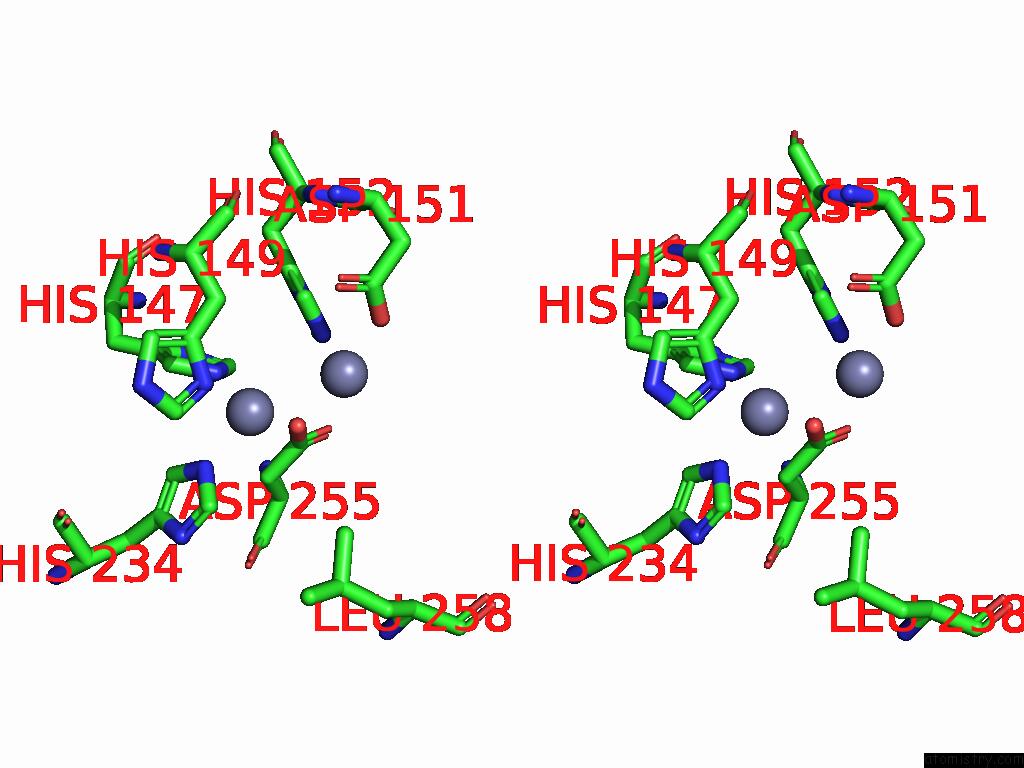
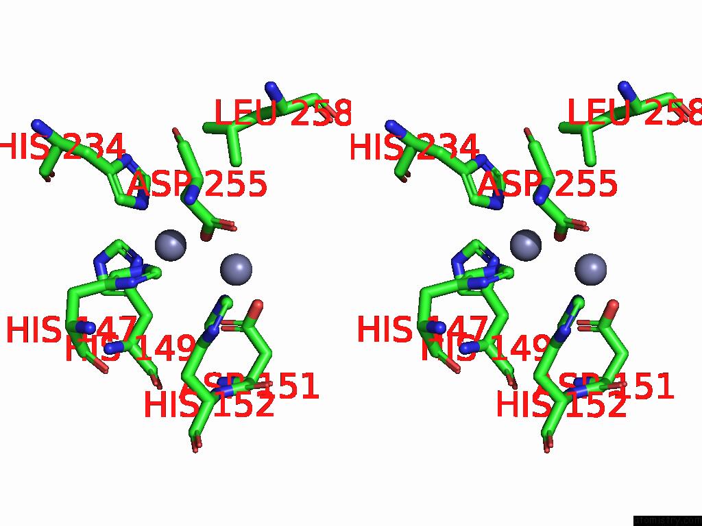
Zinc binding site 2 out
of 4 in 8zu5
Go back to
Zinc Binding Sites List in 8zu5
Zinc binding site 2 out
of 4 in the Crystal Structure of Methyl Parathion Hydrolase Mutant A66D/I143V/I145L/Q272D/S279A/
 Mono view
 Stereo pair view
|
A full contact list of Zinc with other atoms in the Zn binding
site number 2 of Crystal Structure of Methyl Parathion Hydrolase Mutant A66D/I143V/I145L/Q272D/S279A/ within 5.0Å range:
probe
|
atom
|
residue
|
distance (Å)
|
B
|
Occ
|
A:Zn402
b:22.9
occ:1.00
|
OD2
|
A:ASP151
|
2.2
|
18.9
|
1.0
|
NE2
|
A:HIS302
|
2.2
|
17.5
|
1.0
|
OD2
|
A:ASP255
|
2.3
|
18.2
|
1.0
|
NE2
|
A:HIS152
|
2.3
|
15.7
|
1.0
|
CD2
|
A:HIS152
|
2.9
|
16.1
|
1.0
|
CG
|
A:ASP255
|
3.2
|
15.2
|
1.0
|
CE1
|
A:HIS302
|
3.2
|
17.5
|
1.0
|
ZN
|
A:ZN401
|
3.2
|
24.0
|
1.0
|
CG
|
A:ASP151
|
3.2
|
18.8
|
1.0
|
CD2
|
A:HIS302
|
3.3
|
22.2
|
1.0
|
OD1
|
A:ASP255
|
3.3
|
18.3
|
1.0
|
CE1
|
A:HIS152
|
3.5
|
19.0
|
1.0
|
OD1
|
A:ASP151
|
3.6
|
23.1
|
1.0
|
NE2
|
A:HIS147
|
4.0
|
18.1
|
1.0
|
CE1
|
A:HIS147
|
4.2
|
15.4
|
1.0
|
CG
|
A:HIS152
|
4.2
|
16.1
|
1.0
|
ND1
|
A:HIS302
|
4.3
|
20.2
|
1.0
|
CG
|
A:HIS302
|
4.4
|
22.4
|
1.0
|
ND1
|
A:HIS152
|
4.4
|
17.9
|
1.0
|
CB
|
A:ASP151
|
4.5
|
23.0
|
1.0
|
CB
|
A:ASP255
|
4.6
|
18.0
|
1.0
|
CB
|
A:HIS149
|
4.9
|
21.9
|
1.0
|
ND1
|
A:HIS149
|
4.9
|
18.9
|
1.0
|
CD2
|
A:LEU258
|
4.9
|
22.4
|
1.0
|
|
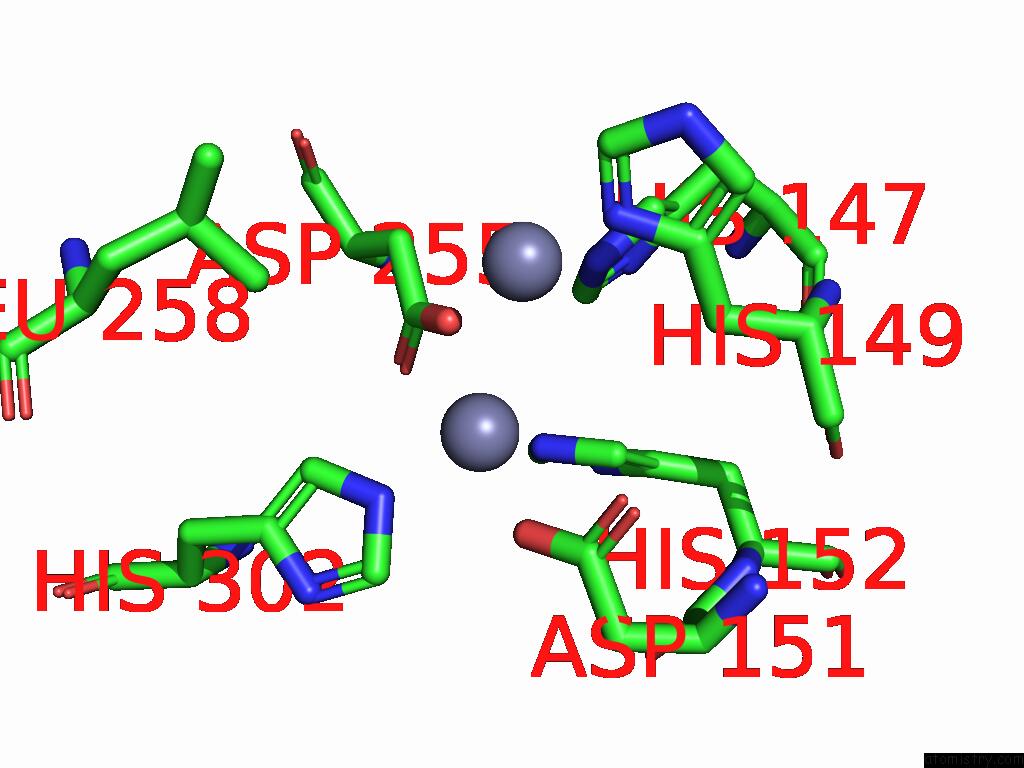
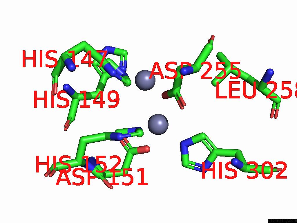
Zinc binding site 3 out
of 4 in 8zu5
Go back to
Zinc Binding Sites List in 8zu5
Zinc binding site 3 out
of 4 in the Crystal Structure of Methyl Parathion Hydrolase Mutant A66D/I143V/I145L/Q272D/S279A/
 Mono view
 Stereo pair view
|
A full contact list of Zinc with other atoms in the Zn binding
site number 3 of Crystal Structure of Methyl Parathion Hydrolase Mutant A66D/I143V/I145L/Q272D/S279A/ within 5.0Å range:
probe
|
atom
|
residue
|
distance (Å)
|
B
|
Occ
|
B:Zn401
b:19.4
occ:1.00
|
OD2
|
B:ASP255
|
2.2
|
15.4
|
1.0
|
NE2
|
B:HIS234
|
2.3
|
12.9
|
1.0
|
ND1
|
B:HIS149
|
2.3
|
15.3
|
1.0
|
NE2
|
B:HIS147
|
2.3
|
12.7
|
1.0
|
CE1
|
B:HIS234
|
3.2
|
19.1
|
1.0
|
ZN
|
B:ZN402
|
3.2
|
18.4
|
1.0
|
CE1
|
B:HIS149
|
3.2
|
20.6
|
1.0
|
CD2
|
B:HIS147
|
3.3
|
13.0
|
1.0
|
CG
|
B:HIS149
|
3.3
|
18.2
|
1.0
|
CE1
|
B:HIS147
|
3.3
|
12.7
|
1.0
|
CG
|
B:ASP255
|
3.3
|
15.2
|
1.0
|
CD2
|
B:HIS234
|
3.3
|
12.9
|
1.0
|
CB
|
B:HIS149
|
3.6
|
15.9
|
1.0
|
CB
|
B:ASP255
|
3.8
|
15.3
|
1.0
|
NE2
|
B:HIS152
|
4.2
|
12.4
|
1.0
|
NE2
|
B:HIS149
|
4.3
|
19.4
|
1.0
|
ND1
|
B:HIS234
|
4.3
|
19.3
|
1.0
|
OD1
|
B:ASP255
|
4.4
|
15.1
|
1.0
|
CD2
|
B:HIS149
|
4.4
|
17.7
|
1.0
|
ND1
|
B:HIS147
|
4.4
|
13.7
|
1.0
|
CG
|
B:HIS147
|
4.4
|
14.4
|
1.0
|
CG
|
B:HIS234
|
4.4
|
15.5
|
1.0
|
CD2
|
B:HIS152
|
4.4
|
13.3
|
1.0
|
OD1
|
B:ASP151
|
4.6
|
17.5
|
1.0
|
OD2
|
B:ASP151
|
4.8
|
14.8
|
1.0
|
CD2
|
B:LEU258
|
4.8
|
20.3
|
1.0
|
|
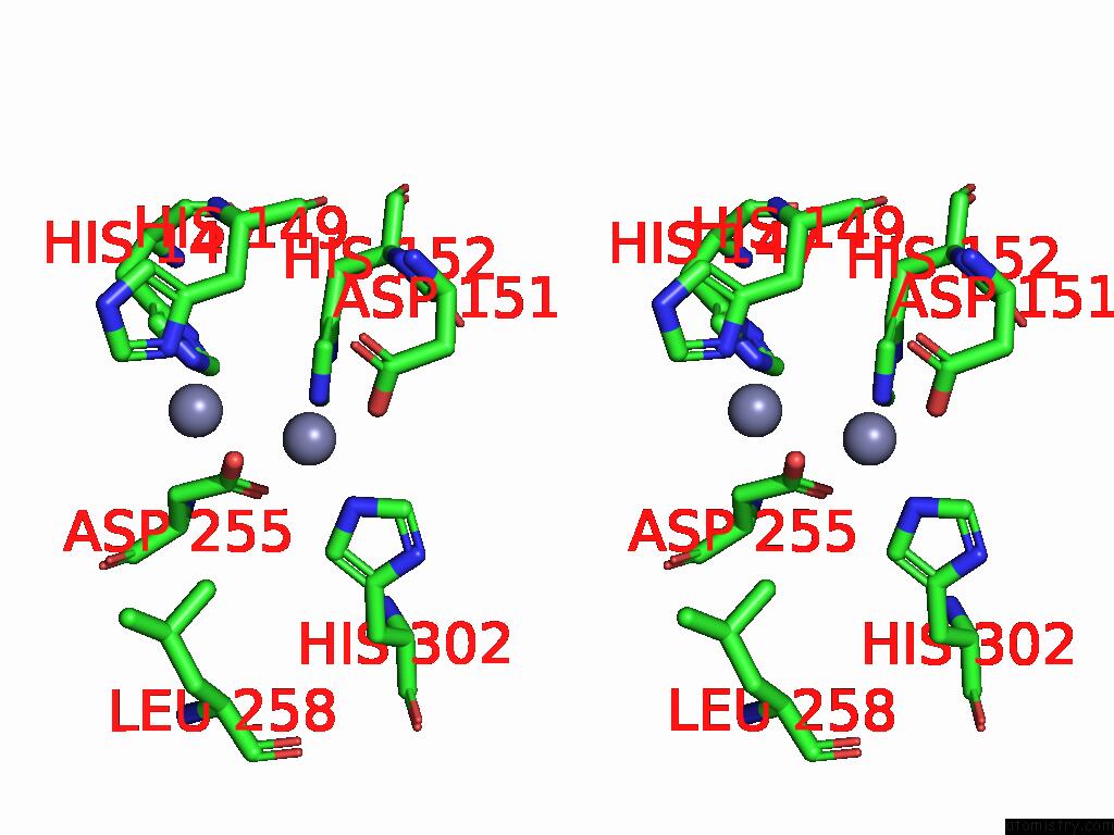
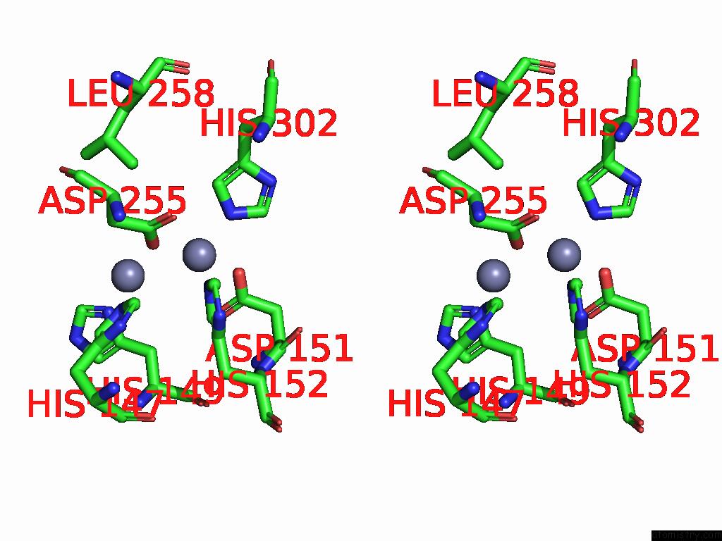
Zinc binding site 4 out
of 4 in 8zu5
Go back to
Zinc Binding Sites List in 8zu5
Zinc binding site 4 out
of 4 in the Crystal Structure of Methyl Parathion Hydrolase Mutant A66D/I143V/I145L/Q272D/S279A/
 Mono view
 Stereo pair view
|
A full contact list of Zinc with other atoms in the Zn binding
site number 4 of Crystal Structure of Methyl Parathion Hydrolase Mutant A66D/I143V/I145L/Q272D/S279A/ within 5.0Å range:
probe
|
atom
|
residue
|
distance (Å)
|
B
|
Occ
|
B:Zn402
b:18.4
occ:1.00
|
OD2
|
B:ASP151
|
2.1
|
14.8
|
1.0
|
OD2
|
B:ASP255
|
2.2
|
15.4
|
1.0
|
NE2
|
B:HIS302
|
2.2
|
15.5
|
1.0
|
NE2
|
B:HIS152
|
2.3
|
12.4
|
1.0
|
CD2
|
B:HIS152
|
3.0
|
13.3
|
1.0
|
CG
|
B:ASP255
|
3.1
|
15.2
|
1.0
|
CG
|
B:ASP151
|
3.1
|
14.9
|
1.0
|
CD2
|
B:HIS302
|
3.1
|
14.4
|
1.0
|
ZN
|
B:ZN401
|
3.2
|
19.4
|
1.0
|
CE1
|
B:HIS302
|
3.2
|
14.7
|
1.0
|
OD1
|
B:ASP255
|
3.3
|
15.1
|
1.0
|
OD1
|
B:ASP151
|
3.4
|
17.5
|
1.0
|
CE1
|
B:HIS152
|
3.4
|
15.9
|
1.0
|
NE2
|
B:HIS147
|
4.1
|
12.7
|
1.0
|
CE1
|
B:HIS147
|
4.2
|
12.7
|
1.0
|
CG
|
B:HIS152
|
4.3
|
12.6
|
1.0
|
CG
|
B:HIS302
|
4.3
|
14.2
|
1.0
|
ND1
|
B:HIS302
|
4.3
|
14.4
|
1.0
|
CB
|
B:ASP151
|
4.4
|
14.2
|
1.0
|
ND1
|
B:HIS152
|
4.4
|
14.5
|
1.0
|
CB
|
B:ASP255
|
4.5
|
15.3
|
1.0
|
CD2
|
B:LEU258
|
4.8
|
20.3
|
1.0
|
CB
|
B:HIS149
|
4.9
|
15.9
|
1.0
|
ND1
|
B:HIS149
|
5.0
|
15.3
|
1.0
|
|
Reference:
F.Xu,
S.Fan,
Y.Li.
Crystal Structure of Methyl Parathion Hydrolase Mutant A66D/I143V/I145L/Q272D/S279A/ To Be Published.
Page generated: Fri Aug 22 16:16:23 2025
|