Zinc in PDB 8ztq: Crystal Structure of Sufu From Mycoplasma Pneumonia
Protein crystallography data
The structure of Crystal Structure of Sufu From Mycoplasma Pneumonia, PDB code: 8ztq
was solved by
W.M.Wang,
D.Y.Ma,
W.J.Gong,
H.Yao,
Y.H.Liu,
H.F.Wang,
with X-Ray Crystallography technique. A brief refinement statistics is given in the table below:
| Resolution Low / High (Å) | 36.78 / 2.89 |
| Space group | C 1 2 1 |
| Cell size a, b, c (Å), α, β, γ (°) | 149.08, 30.333, 87.209, 90, 122.48, 90 |
| R / Rfree (%) | 27.1 / 29.9 |
Zinc Binding Sites:
The binding sites of Zinc atom in the Crystal Structure of Sufu From Mycoplasma Pneumonia
(pdb code 8ztq). This binding sites where shown within
5.0 Angstroms radius around Zinc atom.
In total 2 binding sites of Zinc where determined in the Crystal Structure of Sufu From Mycoplasma Pneumonia, PDB code: 8ztq:
Jump to Zinc binding site number: 1; 2;
In total 2 binding sites of Zinc where determined in the Crystal Structure of Sufu From Mycoplasma Pneumonia, PDB code: 8ztq:
Jump to Zinc binding site number: 1; 2;
Zinc binding site 1 out of 2 in 8ztq
Go back to
Zinc binding site 1 out
of 2 in the Crystal Structure of Sufu From Mycoplasma Pneumonia
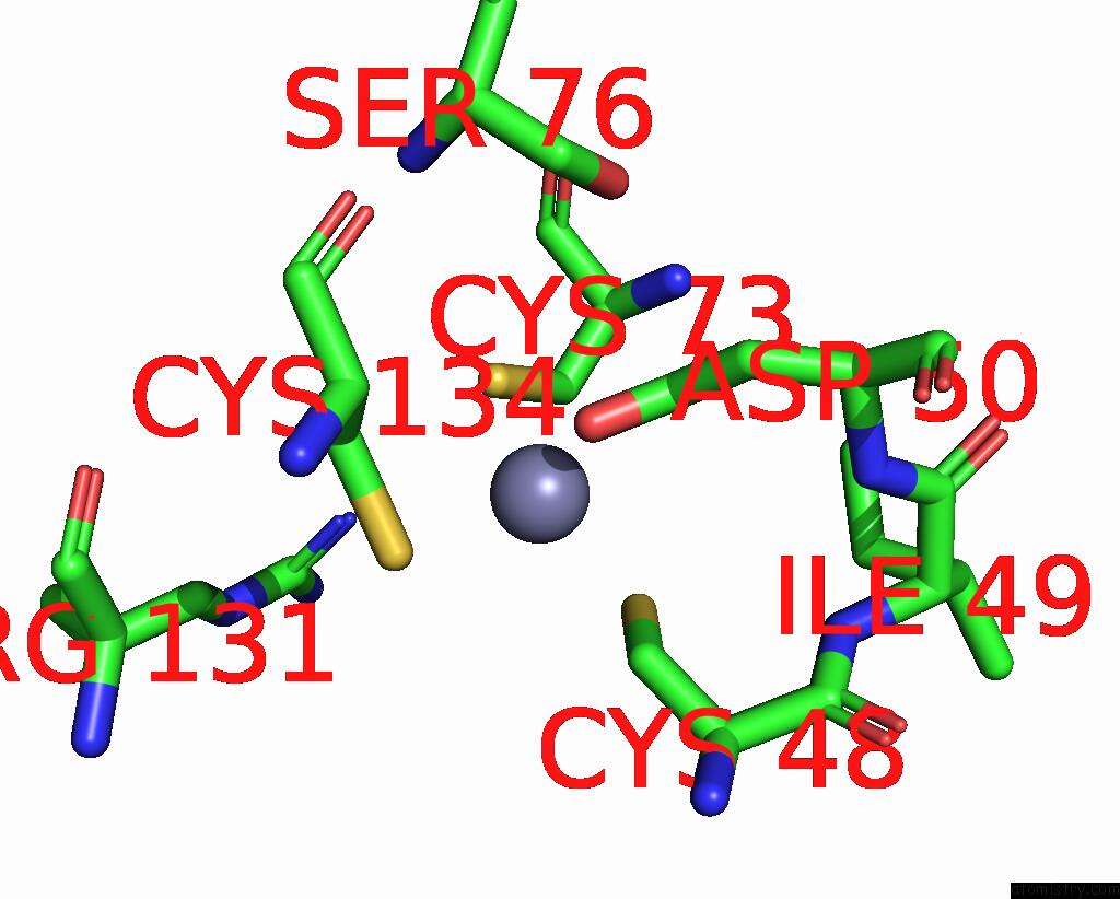
Mono view
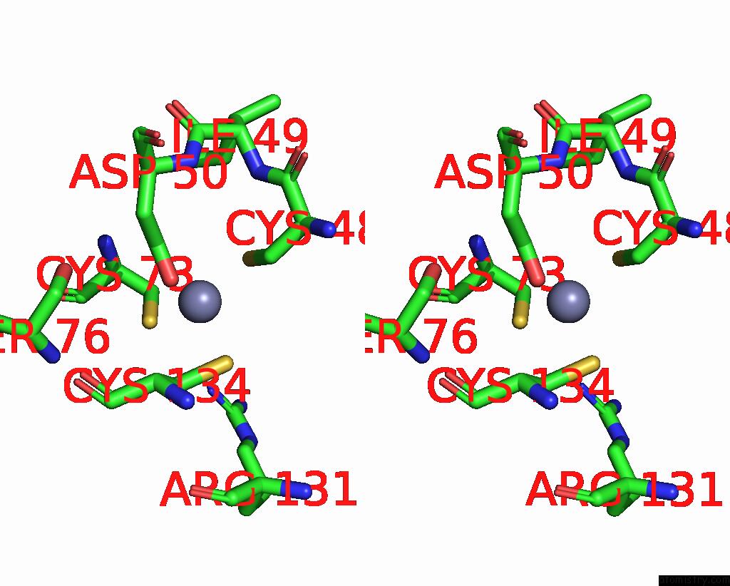
Stereo pair view
Mono view
Stereo pair view
A full contact list of Zinc with other atoms in the Zn binding
site number 1 of Crystal Structure of Sufu From Mycoplasma Pneumonia within 5.0Å range:
|
Zinc binding site 2 out of 2 in 8ztq
Go back to
Zinc binding site 2 out
of 2 in the Crystal Structure of Sufu From Mycoplasma Pneumonia
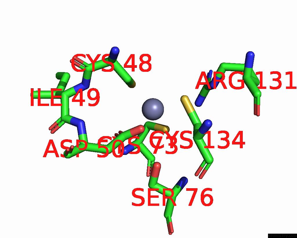
Mono view
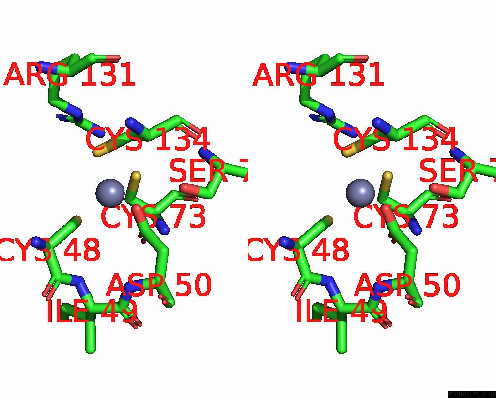
Stereo pair view
Mono view
Stereo pair view
A full contact list of Zinc with other atoms in the Zn binding
site number 2 of Crystal Structure of Sufu From Mycoplasma Pneumonia within 5.0Å range:
|
Reference:
D.Ma,
H.Yao,
Y.Liu,
W.Gong,
Y.Zhao,
R.Wang,
C.Wu,
W.Wang,
H.Wang.
The Reduced Interaction Between Sufs and Sufu in Mycoplasma Penetrans Results in Diminished Sulfotransferase Activity. Int.J.Biol.Macromol. 38181 2024.
ISSN: ISSN 0141-8130
PubMed: 39615726
DOI: 10.1016/J.IJBIOMAC.2024.138181
Page generated: Sun Dec 15 12:15:31 2024
ISSN: ISSN 0141-8130
PubMed: 39615726
DOI: 10.1016/J.IJBIOMAC.2024.138181
Last articles
Zn in 9MJ5Zn in 9HNW
Zn in 9G0L
Zn in 9FNE
Zn in 9DZN
Zn in 9E0I
Zn in 9D32
Zn in 9DAK
Zn in 8ZXC
Zn in 8ZUF