Zinc in PDB 8xzz: Structure of A Xylanase Xyl-1 M4 E175A in Complex with Xylobiose
Enzymatic activity of Structure of A Xylanase Xyl-1 M4 E175A in Complex with Xylobiose
All present enzymatic activity of Structure of A Xylanase Xyl-1 M4 E175A in Complex with Xylobiose:
3.2.1.8;
3.2.1.8;
Protein crystallography data
The structure of Structure of A Xylanase Xyl-1 M4 E175A in Complex with Xylobiose, PDB code: 8xzz
was solved by
W.L.Xiang,
J.-W.Huang,
Y.Yang,
C.-C.Chen,
R.-T.Guo,
with X-Ray Crystallography technique. A brief refinement statistics is given in the table below:
| Resolution Low / High (Å) | 24.27 / 1.72 |
| Space group | P 1 21 1 |
| Cell size a, b, c (Å), α, β, γ (°) | 52.074, 137.54, 52.848, 90, 97.83, 90 |
| R / Rfree (%) | 16.1 / 19.2 |
Zinc Binding Sites:
The binding sites of Zinc atom in the Structure of A Xylanase Xyl-1 M4 E175A in Complex with Xylobiose
(pdb code 8xzz). This binding sites where shown within
5.0 Angstroms radius around Zinc atom.
In total 6 binding sites of Zinc where determined in the Structure of A Xylanase Xyl-1 M4 E175A in Complex with Xylobiose, PDB code: 8xzz:
Jump to Zinc binding site number: 1; 2; 3; 4; 5; 6;
In total 6 binding sites of Zinc where determined in the Structure of A Xylanase Xyl-1 M4 E175A in Complex with Xylobiose, PDB code: 8xzz:
Jump to Zinc binding site number: 1; 2; 3; 4; 5; 6;
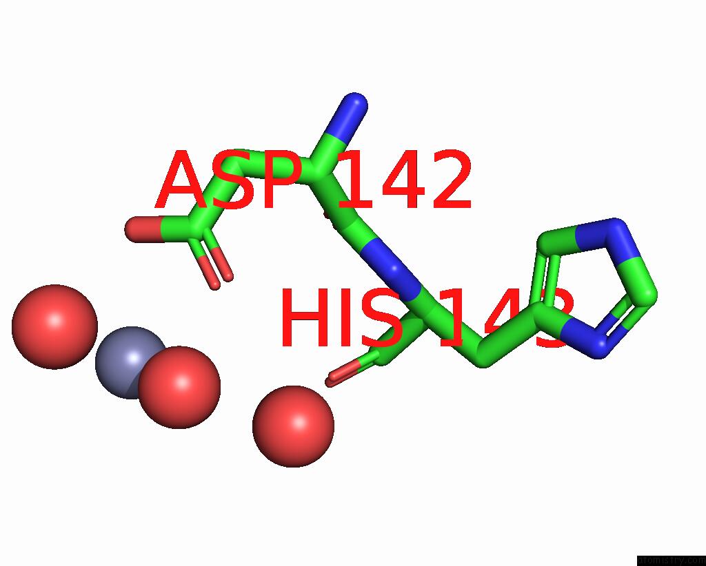
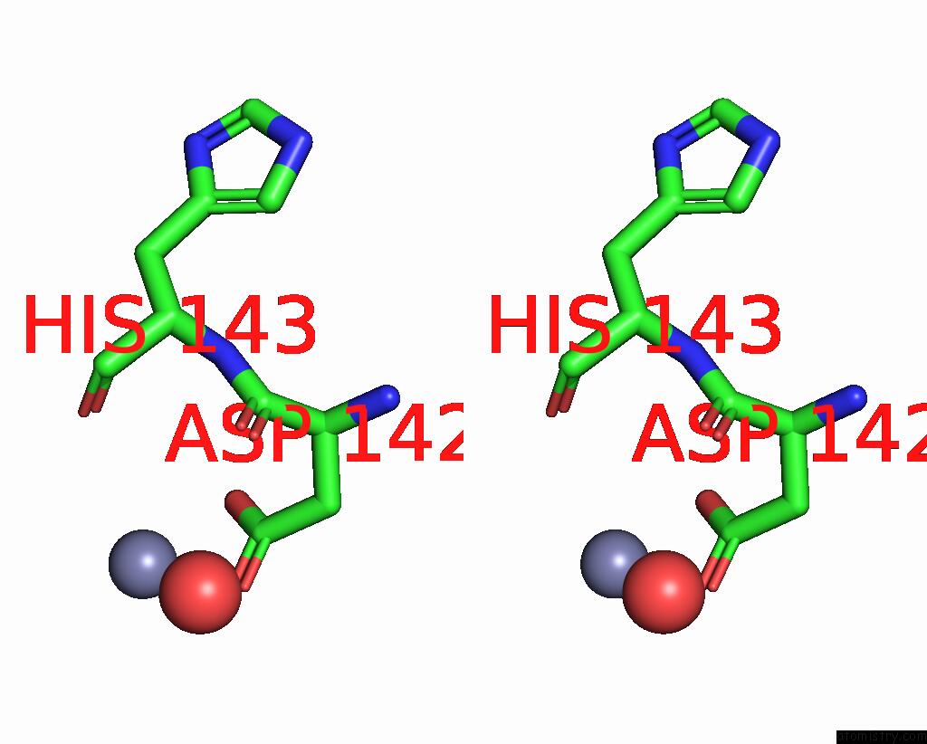
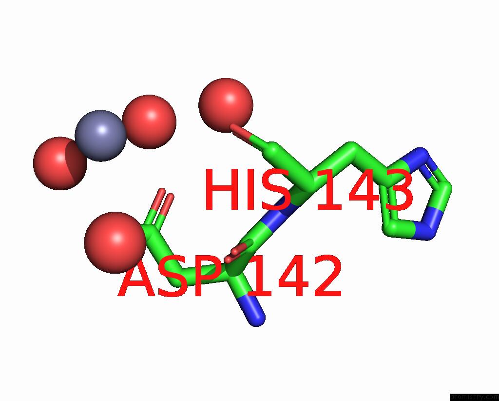
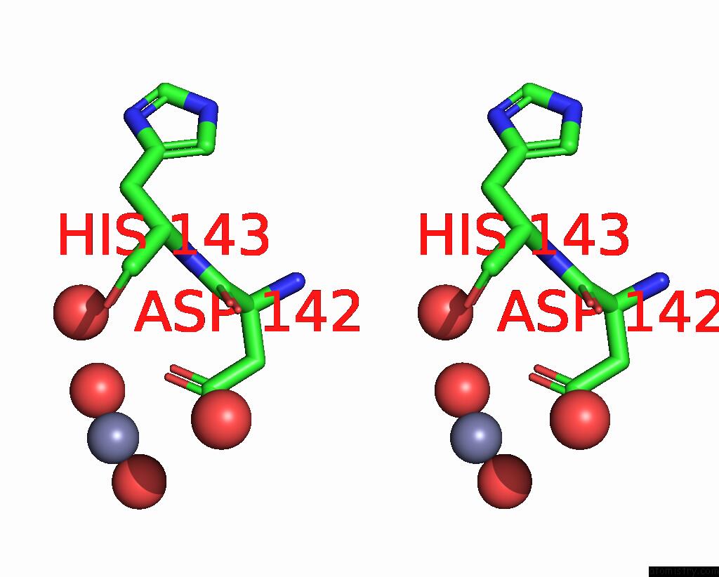
Zinc binding site 1 out of 6 in 8xzz
Go back to
Zinc binding site 1 out
of 6 in the Structure of A Xylanase Xyl-1 M4 E175A in Complex with Xylobiose
Mono view
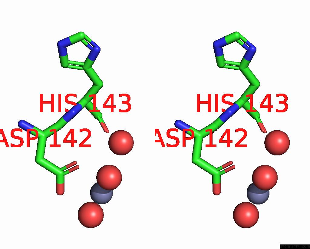
Stereo pair view
Mono view
Stereo pair view
A full contact list of Zinc with other atoms in the Zn binding
site number 1 of Structure of A Xylanase Xyl-1 M4 E175A in Complex with Xylobiose within 5.0Å range:
|
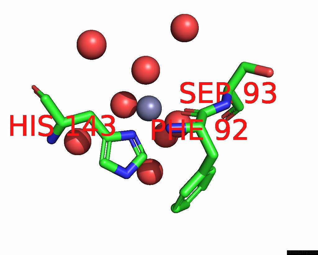
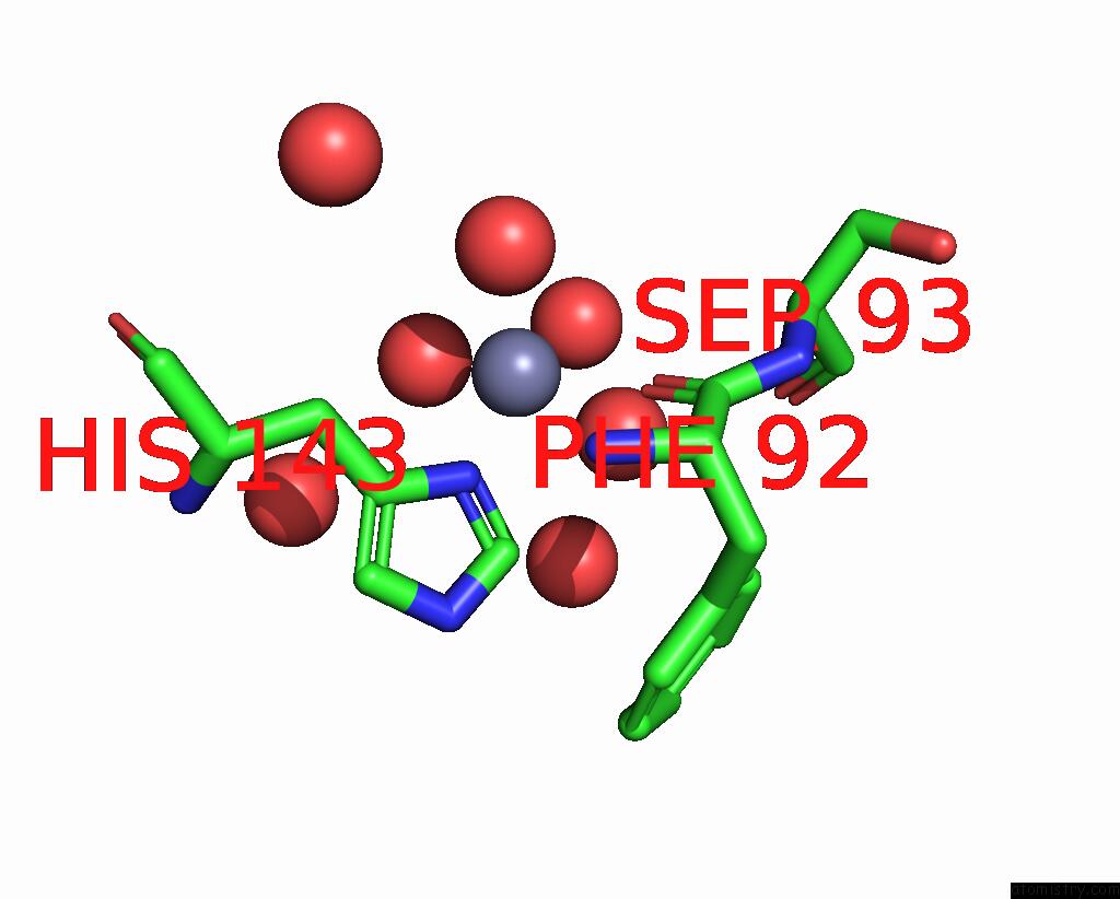
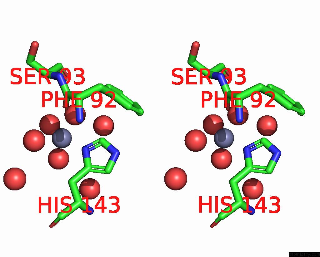
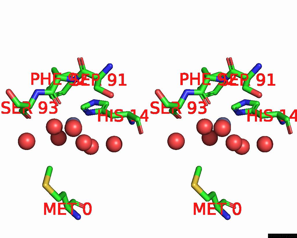
Zinc binding site 2 out of 6 in 8xzz
Go back to
Zinc binding site 2 out
of 6 in the Structure of A Xylanase Xyl-1 M4 E175A in Complex with Xylobiose
Mono view
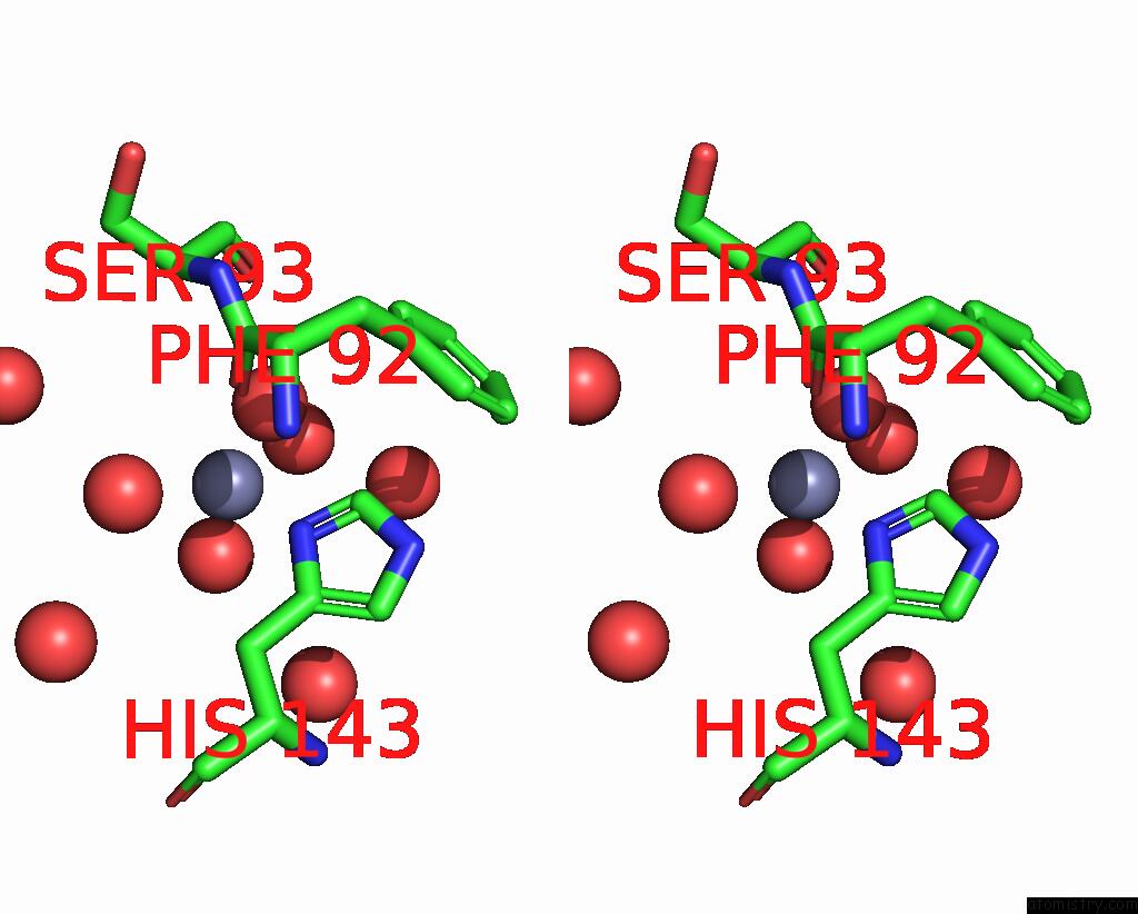
Stereo pair view
Mono view
Stereo pair view
A full contact list of Zinc with other atoms in the Zn binding
site number 2 of Structure of A Xylanase Xyl-1 M4 E175A in Complex with Xylobiose within 5.0Å range:
|
Zinc binding site 3 out of 6 in 8xzz
Go back to
Zinc binding site 3 out
of 6 in the Structure of A Xylanase Xyl-1 M4 E175A in Complex with Xylobiose

Mono view
Stereo pair view
Mono view
Stereo pair view
A full contact list of Zinc with other atoms in the Zn binding
site number 3 of Structure of A Xylanase Xyl-1 M4 E175A in Complex with Xylobiose within 5.0Å range:
|
Zinc binding site 4 out of 6 in 8xzz
Go back to
Zinc binding site 4 out
of 6 in the Structure of A Xylanase Xyl-1 M4 E175A in Complex with Xylobiose
Mono view
Stereo pair view
Mono view
Stereo pair view
A full contact list of Zinc with other atoms in the Zn binding
site number 4 of Structure of A Xylanase Xyl-1 M4 E175A in Complex with Xylobiose within 5.0Å range:
|
Zinc binding site 5 out of 6 in 8xzz
Go back to
Zinc binding site 5 out
of 6 in the Structure of A Xylanase Xyl-1 M4 E175A in Complex with Xylobiose
Mono view
Stereo pair view
Mono view
Stereo pair view
A full contact list of Zinc with other atoms in the Zn binding
site number 5 of Structure of A Xylanase Xyl-1 M4 E175A in Complex with Xylobiose within 5.0Å range:
|
Zinc binding site 6 out of 6 in 8xzz
Go back to
Zinc binding site 6 out
of 6 in the Structure of A Xylanase Xyl-1 M4 E175A in Complex with Xylobiose

Mono view
Stereo pair view
Mono view
Stereo pair view
A full contact list of Zinc with other atoms in the Zn binding
site number 6 of Structure of A Xylanase Xyl-1 M4 E175A in Complex with Xylobiose within 5.0Å range:
|
Reference:
Y.Wu,
Y.Yang,
G.Lu,
W.L.Xiang,
T.Y.Sun,
K.W.Chen,
X.Lv,
Y.F.Gui,
R.Q.Zeng,
Y.K.Du,
C.H.Fu,
J.W.Huang,
C.C.Chen,
R.T.Guo,
L.J.Yu.
Unleashing the Power of Evolution in Xylanase Engineering: Investigating the Role of Distal Mutation Regulation. J.Agric.Food Chem. V. 72 18201 2024.
ISSN: ESSN 1520-5118
PubMed: 39082219
DOI: 10.1021/ACS.JAFC.4C03245
Page generated: Tue Dec 10 21:54:45 2024
ISSN: ESSN 1520-5118
PubMed: 39082219
DOI: 10.1021/ACS.JAFC.4C03245
Last articles
Zn in 9MJ5Zn in 9HNW
Zn in 9G0L
Zn in 9FNE
Zn in 9DZN
Zn in 9E0I
Zn in 9D32
Zn in 9DAK
Zn in 8ZXC
Zn in 8ZUF