Zinc in PDB 8xjg: Crystal Structure of the Yqey Protein From Vibrio Parahaemolyticus
Protein crystallography data
The structure of Crystal Structure of the Yqey Protein From Vibrio Parahaemolyticus, PDB code: 8xjg
was solved by
S.Y.Kim,
S.I.Yoon,
with X-Ray Crystallography technique. A brief refinement statistics is given in the table below:
| Resolution Low / High (Å) | 29.14 / 1.70 |
| Space group | P 21 21 2 |
| Cell size a, b, c (Å), α, β, γ (°) | 43.721, 107.577, 34.671, 90, 90, 90 |
| R / Rfree (%) | 22.7 / 25.8 |
Zinc Binding Sites:
The binding sites of Zinc atom in the Crystal Structure of the Yqey Protein From Vibrio Parahaemolyticus
(pdb code 8xjg). This binding sites where shown within
5.0 Angstroms radius around Zinc atom.
In total 3 binding sites of Zinc where determined in the Crystal Structure of the Yqey Protein From Vibrio Parahaemolyticus, PDB code: 8xjg:
Jump to Zinc binding site number: 1; 2; 3;
In total 3 binding sites of Zinc where determined in the Crystal Structure of the Yqey Protein From Vibrio Parahaemolyticus, PDB code: 8xjg:
Jump to Zinc binding site number: 1; 2; 3;
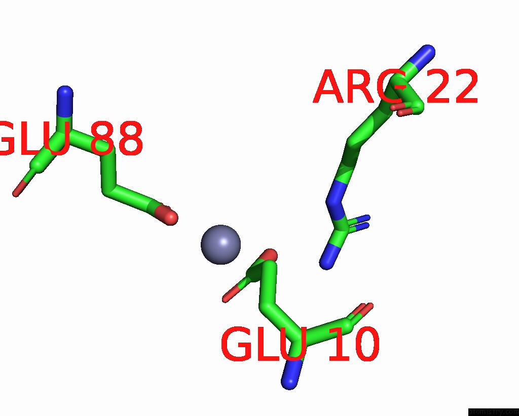
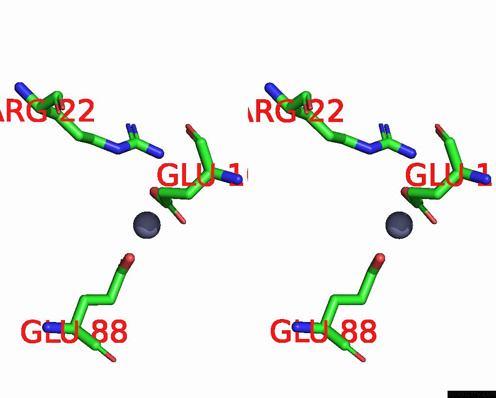
Zinc binding site 1 out of 3 in 8xjg
Go back to
Zinc binding site 1 out
of 3 in the Crystal Structure of the Yqey Protein From Vibrio Parahaemolyticus
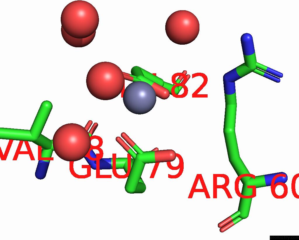
Mono view
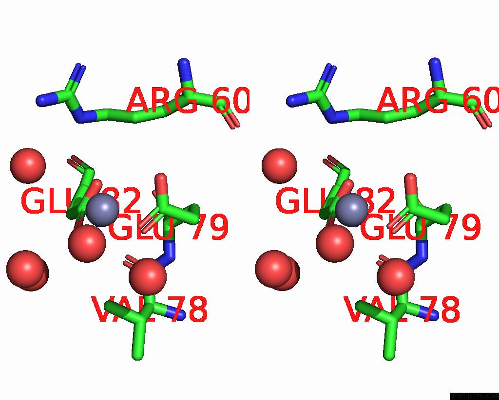
Stereo pair view
Mono view
Stereo pair view
A full contact list of Zinc with other atoms in the Zn binding
site number 1 of Crystal Structure of the Yqey Protein From Vibrio Parahaemolyticus within 5.0Å range:
|
Zinc binding site 2 out of 3 in 8xjg
Go back to
Zinc binding site 2 out
of 3 in the Crystal Structure of the Yqey Protein From Vibrio Parahaemolyticus
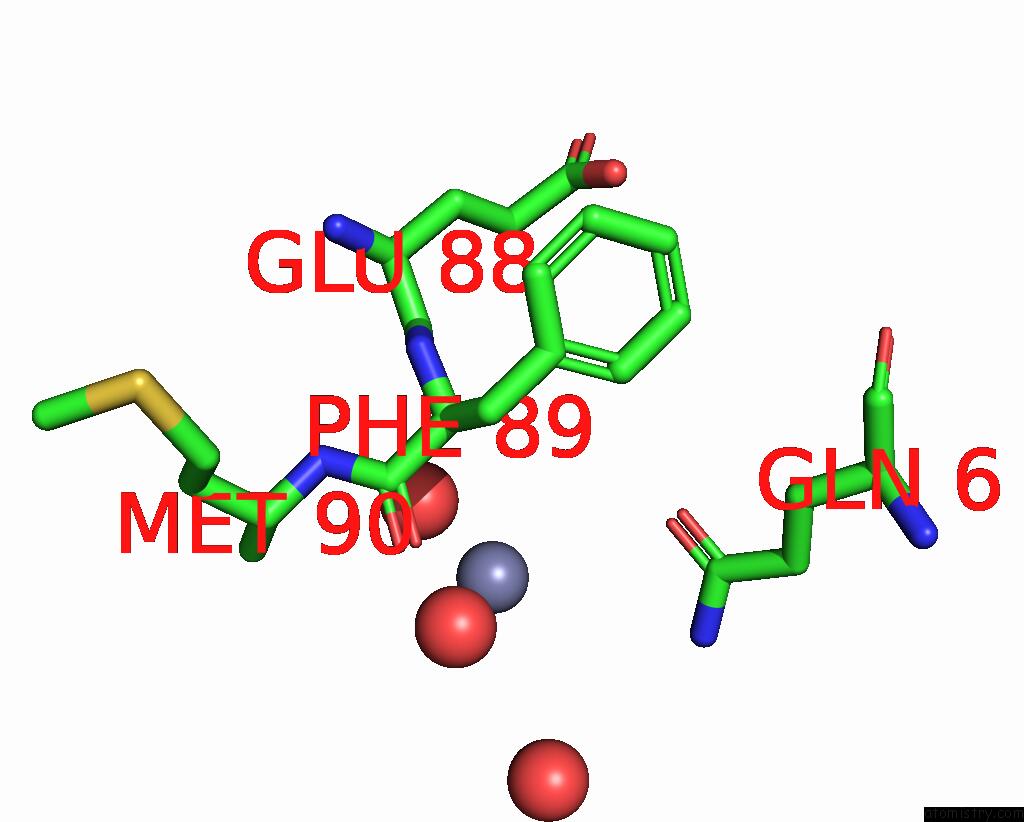
Mono view
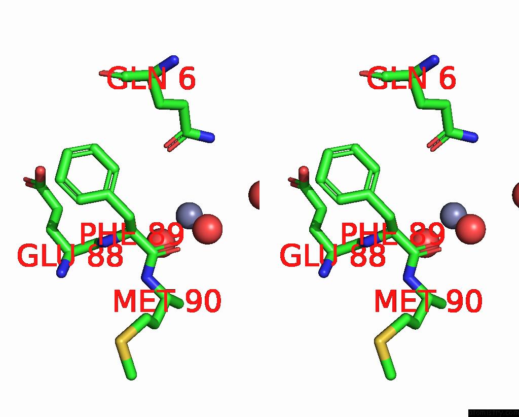
Stereo pair view
Mono view
Stereo pair view
A full contact list of Zinc with other atoms in the Zn binding
site number 2 of Crystal Structure of the Yqey Protein From Vibrio Parahaemolyticus within 5.0Å range:
|
Zinc binding site 3 out of 3 in 8xjg
Go back to
Zinc binding site 3 out
of 3 in the Crystal Structure of the Yqey Protein From Vibrio Parahaemolyticus
Mono view
Stereo pair view
Mono view
Stereo pair view
A full contact list of Zinc with other atoms in the Zn binding
site number 3 of Crystal Structure of the Yqey Protein From Vibrio Parahaemolyticus within 5.0Å range:
|
Reference:
S.Y.Kim,
S.I.Yoon.
Structural Analysis of the Yqey Proteins From Campylobacter Jejuni and Vibrio Parahaemolyticus. Biochem.Biophys.Res.Commun. V. 695 49485 2024.
ISSN: ESSN 1090-2104
PubMed: 38211535
DOI: 10.1016/J.BBRC.2024.149485
Page generated: Thu Oct 31 13:53:50 2024
ISSN: ESSN 1090-2104
PubMed: 38211535
DOI: 10.1016/J.BBRC.2024.149485
Last articles
Zn in 9MJ5Zn in 9HNW
Zn in 9G0L
Zn in 9FNE
Zn in 9DZN
Zn in 9E0I
Zn in 9D32
Zn in 9DAK
Zn in 8ZXC
Zn in 8ZUF