Zinc in PDB 8xf8: High-Resolution Structure of the Siderophore Periplasmic Binding Protein Ftsb From Streptococcus Pyogenes with Ferrioxamine B
Protein crystallography data
The structure of High-Resolution Structure of the Siderophore Periplasmic Binding Protein Ftsb From Streptococcus Pyogenes with Ferrioxamine B, PDB code: 8xf8
was solved by
J.M.M.Caaveiro,
J.Fernandez-Perez,
K.Tsumoto,
with X-Ray Crystallography technique. A brief refinement statistics is given in the table below:
| Resolution Low / High (Å) | 40.50 / 1.15 |
| Space group | P 65 |
| Cell size a, b, c (Å), α, β, γ (°) | 76.223, 76.223, 102.502, 90, 90, 120 |
| R / Rfree (%) | 13.5 / 15.3 |
Other elements in 8xf8:
The structure of High-Resolution Structure of the Siderophore Periplasmic Binding Protein Ftsb From Streptococcus Pyogenes with Ferrioxamine B also contains other interesting chemical elements:
| Sodium | (Na) | 2 atoms |
| Iron | (Fe) | 1 atom |
Zinc Binding Sites:
The binding sites of Zinc atom in the High-Resolution Structure of the Siderophore Periplasmic Binding Protein Ftsb From Streptococcus Pyogenes with Ferrioxamine B
(pdb code 8xf8). This binding sites where shown within
5.0 Angstroms radius around Zinc atom.
In total 5 binding sites of Zinc where determined in the High-Resolution Structure of the Siderophore Periplasmic Binding Protein Ftsb From Streptococcus Pyogenes with Ferrioxamine B, PDB code: 8xf8:
Jump to Zinc binding site number: 1; 2; 3; 4; 5;
In total 5 binding sites of Zinc where determined in the High-Resolution Structure of the Siderophore Periplasmic Binding Protein Ftsb From Streptococcus Pyogenes with Ferrioxamine B, PDB code: 8xf8:
Jump to Zinc binding site number: 1; 2; 3; 4; 5;
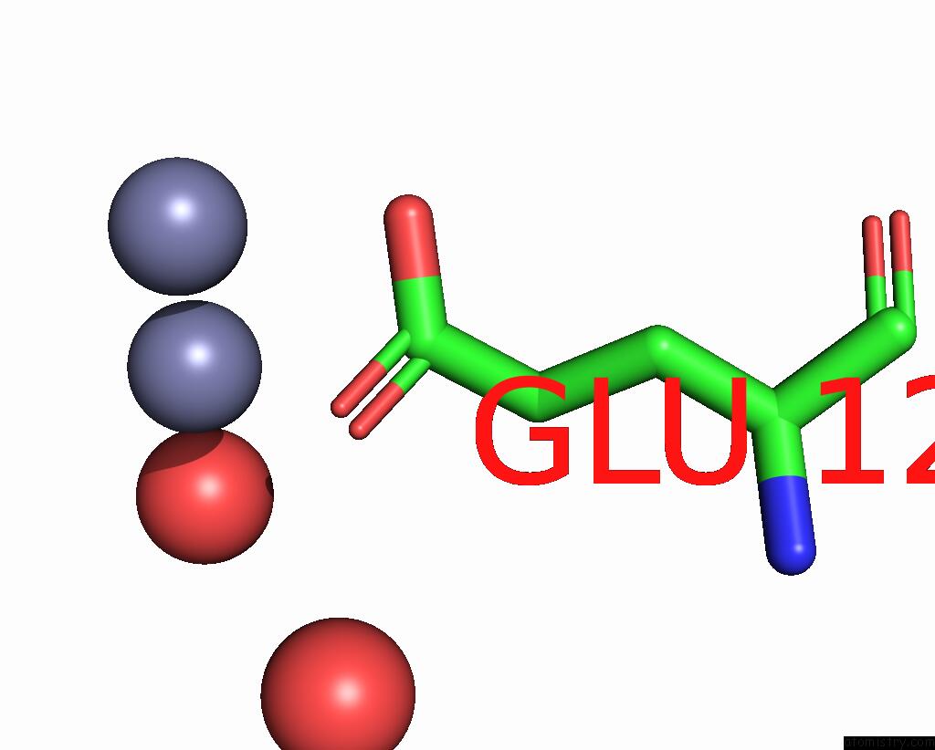
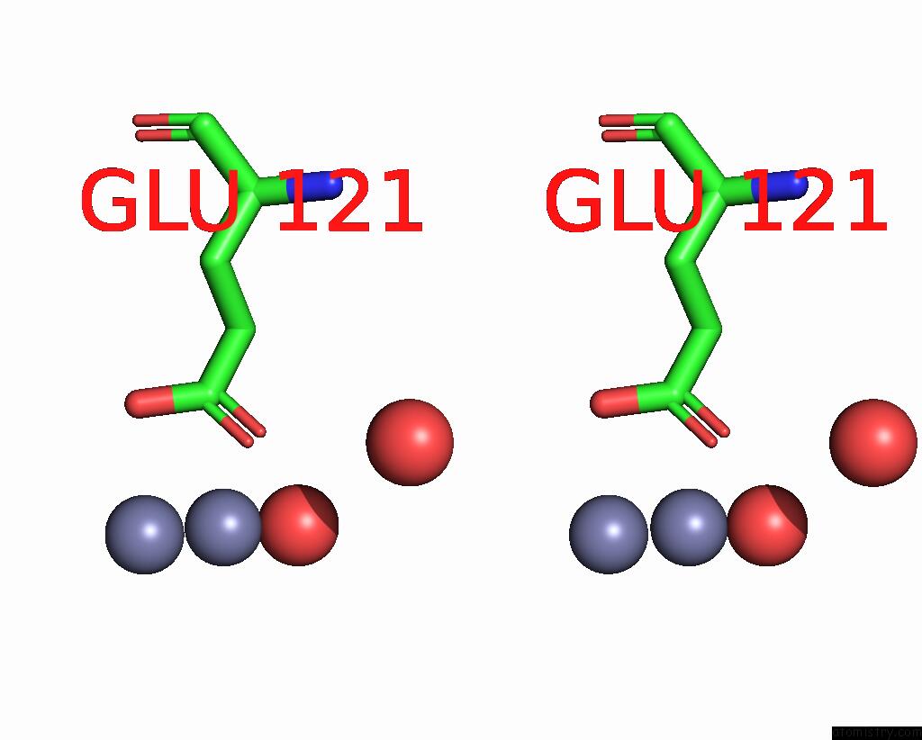
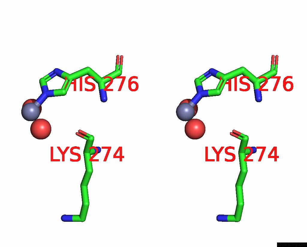
Zinc binding site 1 out of 5 in 8xf8
Go back to
Zinc binding site 1 out
of 5 in the High-Resolution Structure of the Siderophore Periplasmic Binding Protein Ftsb From Streptococcus Pyogenes with Ferrioxamine B
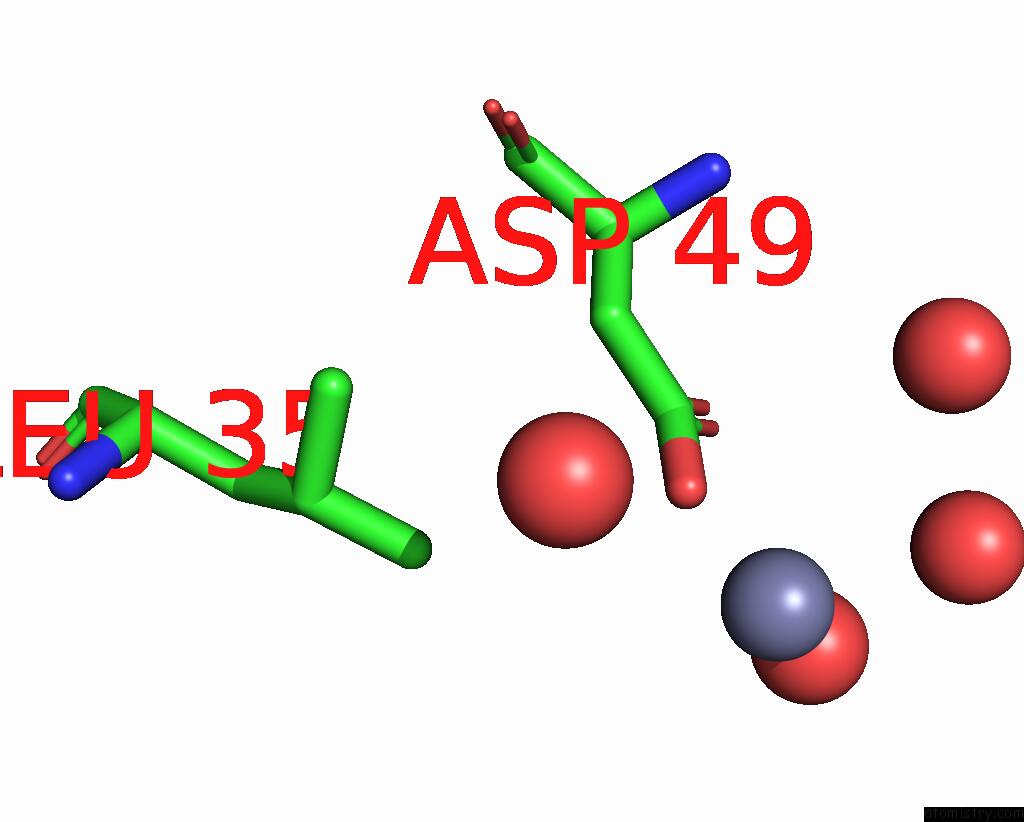
Mono view
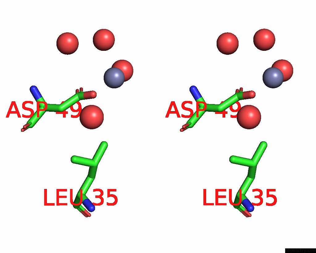
Stereo pair view
Mono view
Stereo pair view
A full contact list of Zinc with other atoms in the Zn binding
site number 1 of High-Resolution Structure of the Siderophore Periplasmic Binding Protein Ftsb From Streptococcus Pyogenes with Ferrioxamine B within 5.0Å range:
|
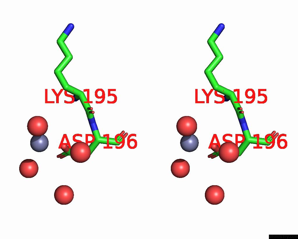
Zinc binding site 2 out of 5 in 8xf8
Go back to
Zinc binding site 2 out
of 5 in the High-Resolution Structure of the Siderophore Periplasmic Binding Protein Ftsb From Streptococcus Pyogenes with Ferrioxamine B
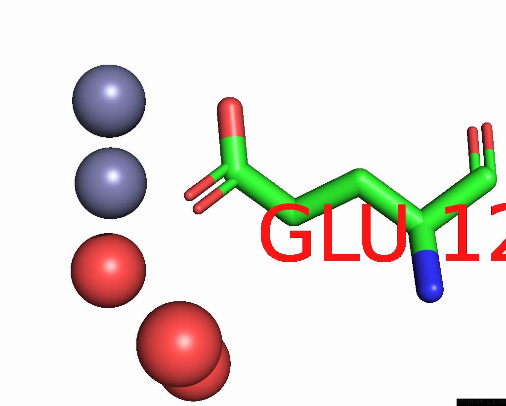
Mono view
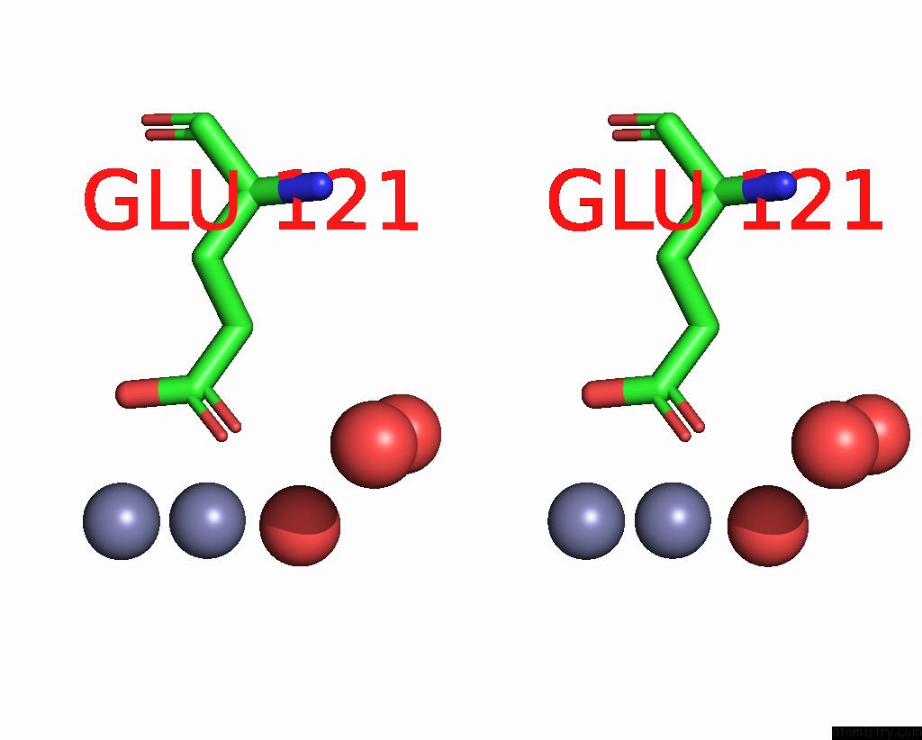
Stereo pair view
Mono view
Stereo pair view
A full contact list of Zinc with other atoms in the Zn binding
site number 2 of High-Resolution Structure of the Siderophore Periplasmic Binding Protein Ftsb From Streptococcus Pyogenes with Ferrioxamine B within 5.0Å range:
|
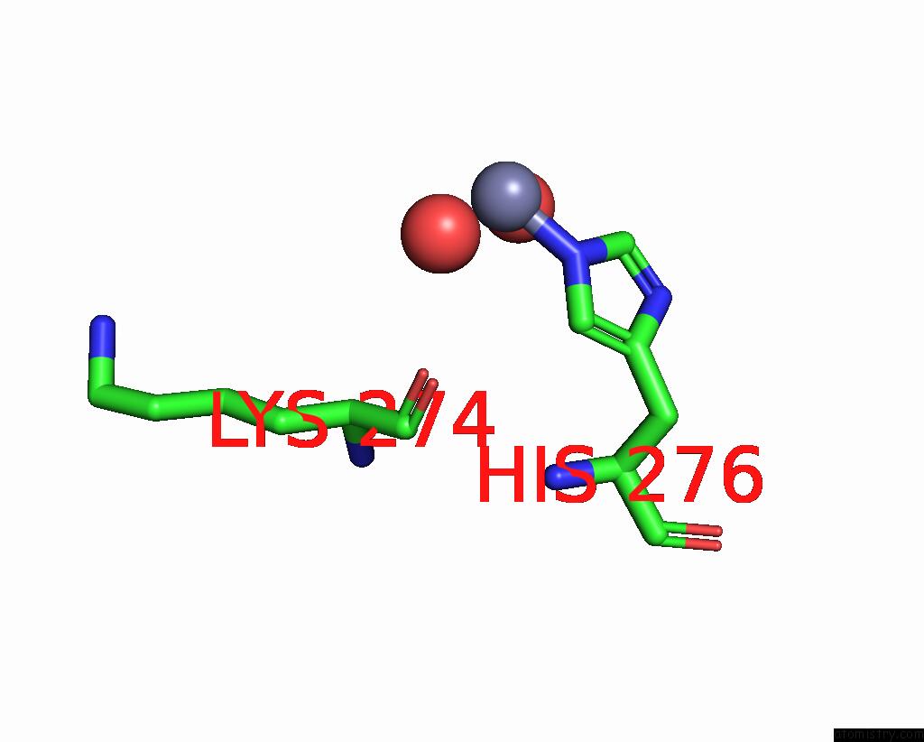
Zinc binding site 3 out of 5 in 8xf8
Go back to
Zinc binding site 3 out
of 5 in the High-Resolution Structure of the Siderophore Periplasmic Binding Protein Ftsb From Streptococcus Pyogenes with Ferrioxamine B
Mono view
Stereo pair view
Mono view
Stereo pair view
A full contact list of Zinc with other atoms in the Zn binding
site number 3 of High-Resolution Structure of the Siderophore Periplasmic Binding Protein Ftsb From Streptococcus Pyogenes with Ferrioxamine B within 5.0Å range:
|
Zinc binding site 4 out of 5 in 8xf8
Go back to
Zinc binding site 4 out
of 5 in the High-Resolution Structure of the Siderophore Periplasmic Binding Protein Ftsb From Streptococcus Pyogenes with Ferrioxamine B

Mono view
Stereo pair view
Mono view
Stereo pair view
A full contact list of Zinc with other atoms in the Zn binding
site number 4 of High-Resolution Structure of the Siderophore Periplasmic Binding Protein Ftsb From Streptococcus Pyogenes with Ferrioxamine B within 5.0Å range:
|
Zinc binding site 5 out of 5 in 8xf8
Go back to
Zinc binding site 5 out
of 5 in the High-Resolution Structure of the Siderophore Periplasmic Binding Protein Ftsb From Streptococcus Pyogenes with Ferrioxamine B
Mono view
Stereo pair view
Mono view
Stereo pair view
A full contact list of Zinc with other atoms in the Zn binding
site number 5 of High-Resolution Structure of the Siderophore Periplasmic Binding Protein Ftsb From Streptococcus Pyogenes with Ferrioxamine B within 5.0Å range:
|
Reference:
J.Fernandez-Perez,
J.M.M.Caaveiro,
A.Senoo,
M.Nakakido,
S.De Vega,
I.Nakagawa,
K.Tsumoto.
Conserved Binding Mechanism For Ligand Promiscuity in the Hydroxamate Siderophore Binding Protein Ftsb From Streptococcus Pyogenes Structure.
ISSN: ISSN 0969-2126
DOI: 10.1016/J.STR.2024.09.018
Page generated: Thu Oct 31 13:53:56 2024
ISSN: ISSN 0969-2126
DOI: 10.1016/J.STR.2024.09.018
Last articles
Zn in 9MJ5Zn in 9HNW
Zn in 9G0L
Zn in 9FNE
Zn in 9DZN
Zn in 9E0I
Zn in 9D32
Zn in 9DAK
Zn in 8ZXC
Zn in 8ZUF