Zinc »
PDB 8x3w-8xld »
8x4p »
Zinc in PDB 8x4p: Crystal Structure of the Y135A Mutant of DIMT1 in Complex with Adenosylornithine (Sfg) From Pyrococcus Horikoshii
Enzymatic activity of Crystal Structure of the Y135A Mutant of DIMT1 in Complex with Adenosylornithine (Sfg) From Pyrococcus Horikoshii
All present enzymatic activity of Crystal Structure of the Y135A Mutant of DIMT1 in Complex with Adenosylornithine (Sfg) From Pyrococcus Horikoshii:
2.1.1.182;
2.1.1.182;
Protein crystallography data
The structure of Crystal Structure of the Y135A Mutant of DIMT1 in Complex with Adenosylornithine (Sfg) From Pyrococcus Horikoshii, PDB code: 8x4p
was solved by
S.Saha,
S.P.Kanaujia,
with X-Ray Crystallography technique. A brief refinement statistics is given in the table below:
| Resolution Low / High (Å) | 76.58 / 2.60 |
| Space group | C 1 2 1 |
| Cell size a, b, c (Å), α, β, γ (°) | 119.06, 79.96, 85.41, 90, 116.28, 90 |
| R / Rfree (%) | 26.2 / 28.9 |
Zinc Binding Sites:
The binding sites of Zinc atom in the Crystal Structure of the Y135A Mutant of DIMT1 in Complex with Adenosylornithine (Sfg) From Pyrococcus Horikoshii
(pdb code 8x4p). This binding sites where shown within
5.0 Angstroms radius around Zinc atom.
In total 4 binding sites of Zinc where determined in the Crystal Structure of the Y135A Mutant of DIMT1 in Complex with Adenosylornithine (Sfg) From Pyrococcus Horikoshii, PDB code: 8x4p:
Jump to Zinc binding site number: 1; 2; 3; 4;
In total 4 binding sites of Zinc where determined in the Crystal Structure of the Y135A Mutant of DIMT1 in Complex with Adenosylornithine (Sfg) From Pyrococcus Horikoshii, PDB code: 8x4p:
Jump to Zinc binding site number: 1; 2; 3; 4;
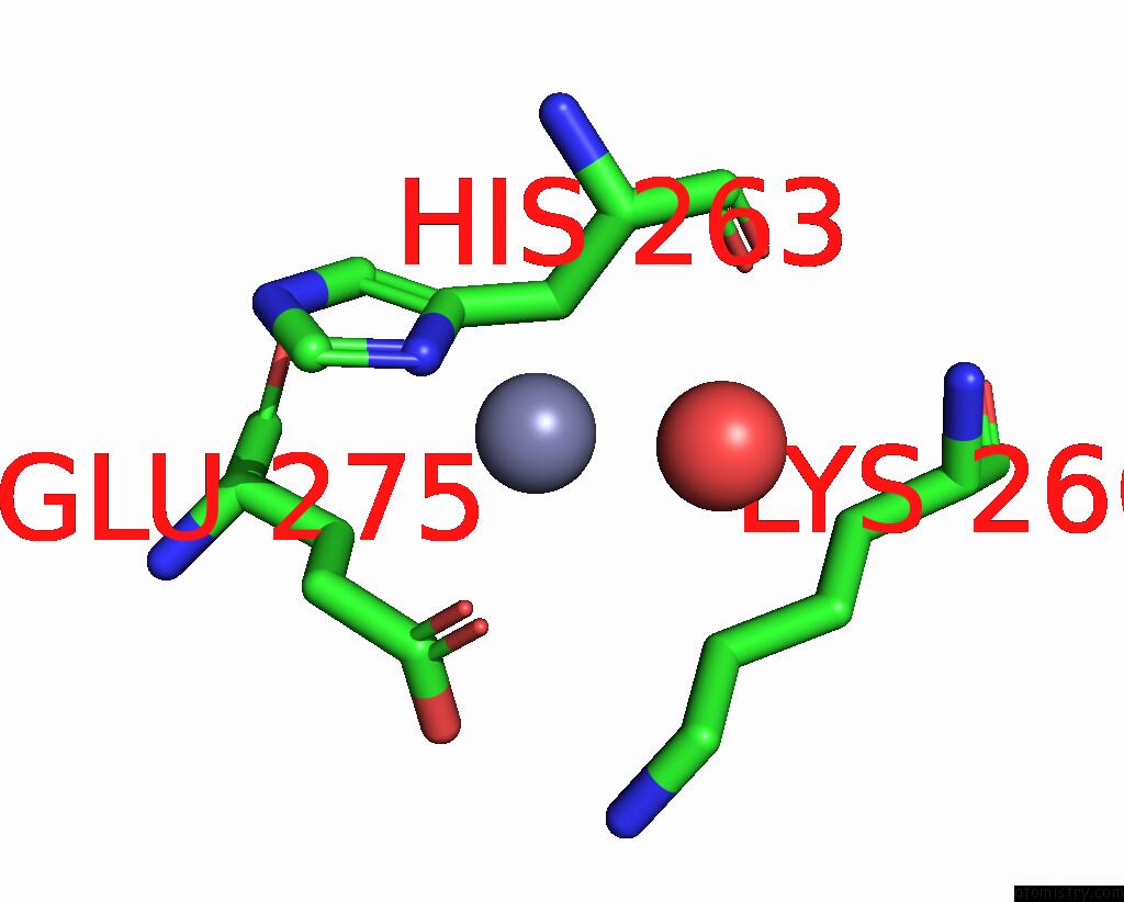
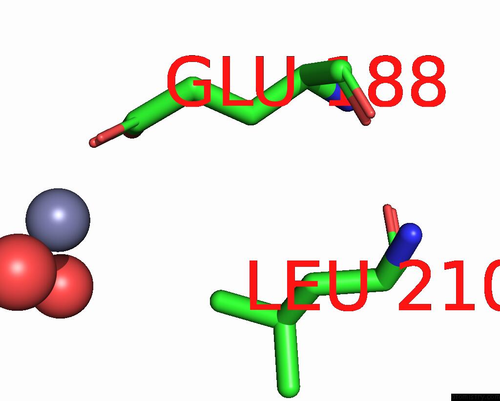
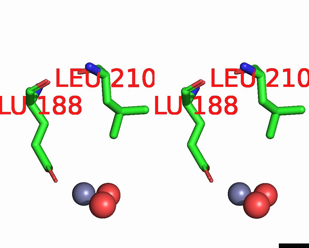
Zinc binding site 1 out of 4 in 8x4p
Go back to
Zinc binding site 1 out
of 4 in the Crystal Structure of the Y135A Mutant of DIMT1 in Complex with Adenosylornithine (Sfg) From Pyrococcus Horikoshii
Mono view
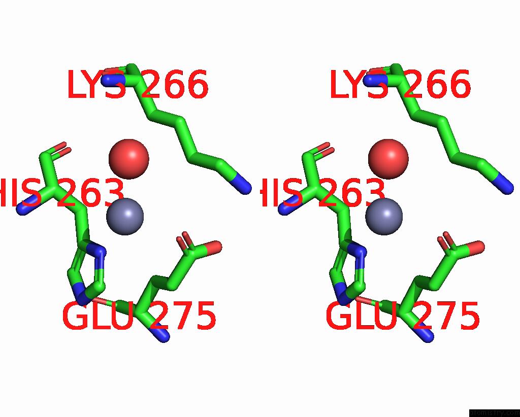
Stereo pair view
Mono view
Stereo pair view
A full contact list of Zinc with other atoms in the Zn binding
site number 1 of Crystal Structure of the Y135A Mutant of DIMT1 in Complex with Adenosylornithine (Sfg) From Pyrococcus Horikoshii within 5.0Å range:
|
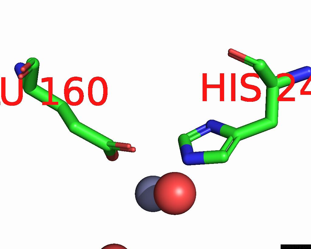
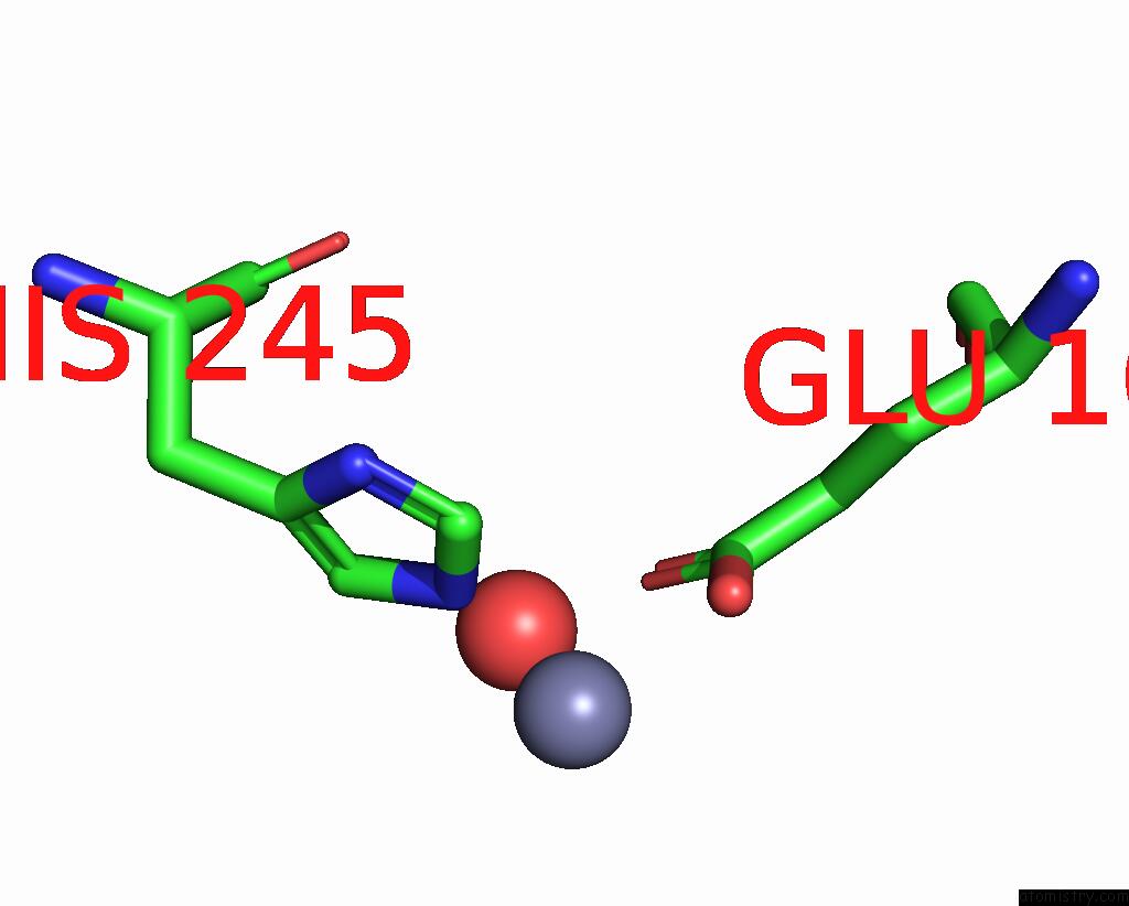
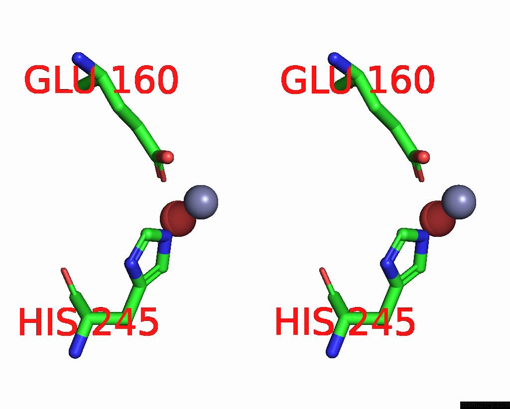
Zinc binding site 2 out of 4 in 8x4p
Go back to
Zinc binding site 2 out
of 4 in the Crystal Structure of the Y135A Mutant of DIMT1 in Complex with Adenosylornithine (Sfg) From Pyrococcus Horikoshii
Mono view
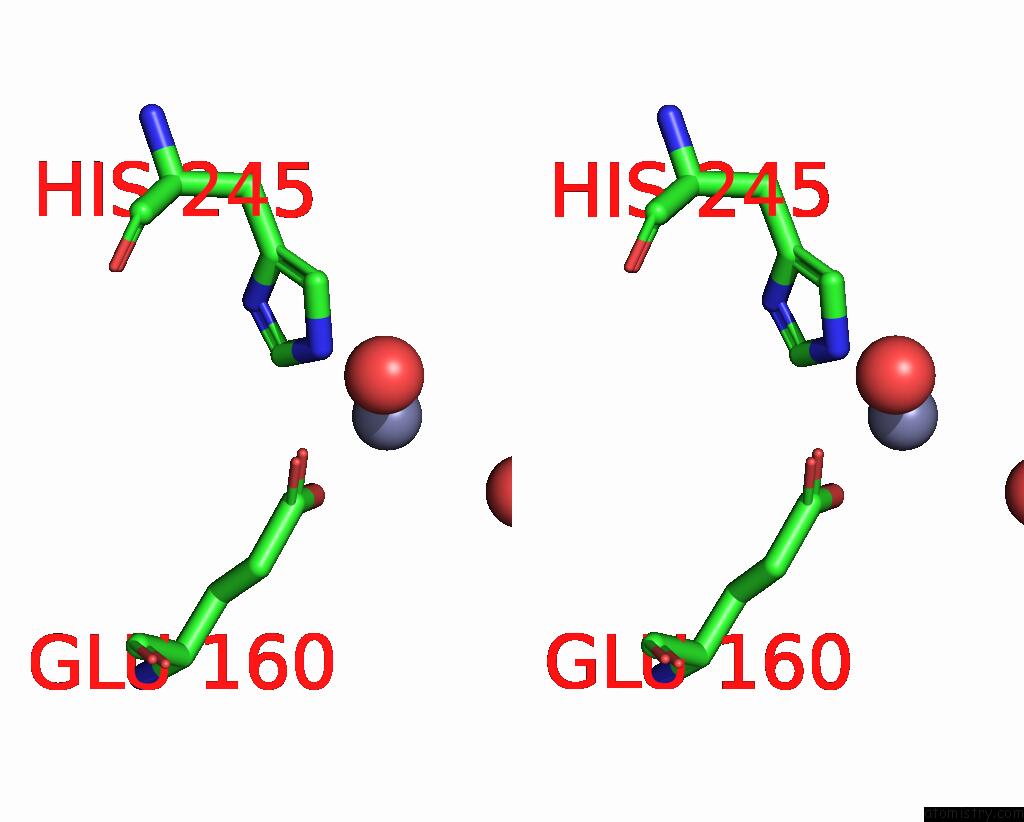
Stereo pair view
Mono view
Stereo pair view
A full contact list of Zinc with other atoms in the Zn binding
site number 2 of Crystal Structure of the Y135A Mutant of DIMT1 in Complex with Adenosylornithine (Sfg) From Pyrococcus Horikoshii within 5.0Å range:
|
Zinc binding site 3 out of 4 in 8x4p
Go back to
Zinc binding site 3 out
of 4 in the Crystal Structure of the Y135A Mutant of DIMT1 in Complex with Adenosylornithine (Sfg) From Pyrococcus Horikoshii
Mono view
Stereo pair view
Mono view
Stereo pair view
A full contact list of Zinc with other atoms in the Zn binding
site number 3 of Crystal Structure of the Y135A Mutant of DIMT1 in Complex with Adenosylornithine (Sfg) From Pyrococcus Horikoshii within 5.0Å range:
|
Zinc binding site 4 out of 4 in 8x4p
Go back to
Zinc binding site 4 out
of 4 in the Crystal Structure of the Y135A Mutant of DIMT1 in Complex with Adenosylornithine (Sfg) From Pyrococcus Horikoshii
Mono view
Stereo pair view
Mono view
Stereo pair view
A full contact list of Zinc with other atoms in the Zn binding
site number 4 of Crystal Structure of the Y135A Mutant of DIMT1 in Complex with Adenosylornithine (Sfg) From Pyrococcus Horikoshii within 5.0Å range:
|
Reference:
S.Saha,
S.P.Kanaujia.
Structural and Functional Characterization of Archaeal DIMT1 Unveils Distinct Protein Dynamics Essential For Efficient Catalysis. Structure 2024.
ISSN: ISSN 0969-2126
PubMed: 39146930
DOI: 10.1016/J.STR.2024.07.013
Page generated: Fri Aug 22 15:25:12 2025
ISSN: ISSN 0969-2126
PubMed: 39146930
DOI: 10.1016/J.STR.2024.07.013
Last articles
Zr in 1XC1Zr in 6Y7P
Zr in 6GNL
Zr in 6HYB
Zr in 4XYY
Zr in 5KHP
Zn in 9VXG
Zn in 9VWY
Zn in 9VCL
Zn in 9VKN