Zinc »
PDB 8we0-8wo3 »
8wig »
Zinc in PDB 8wig: Crystal Structure of E. Coli Thrs Catalytic Domain Mutant G463S/Q484A
Enzymatic activity of Crystal Structure of E. Coli Thrs Catalytic Domain Mutant G463S/Q484A
All present enzymatic activity of Crystal Structure of E. Coli Thrs Catalytic Domain Mutant G463S/Q484A:
6.1.1.3;
6.1.1.3;
Protein crystallography data
The structure of Crystal Structure of E. Coli Thrs Catalytic Domain Mutant G463S/Q484A, PDB code: 8wig
was solved by
H.Qiao,
Z.Wang,
J.Wang,
P.Fang,
with X-Ray Crystallography technique. A brief refinement statistics is given in the table below:
| Resolution Low / High (Å) | 56.21 / 3.22 |
| Space group | P 21 21 21 |
| Cell size a, b, c (Å), α, β, γ (°) | 90.127, 108.179, 112.418, 90, 90, 90 |
| R / Rfree (%) | 20.8 / 22.2 |
Zinc Binding Sites:
The binding sites of Zinc atom in the Crystal Structure of E. Coli Thrs Catalytic Domain Mutant G463S/Q484A
(pdb code 8wig). This binding sites where shown within
5.0 Angstroms radius around Zinc atom.
In total 2 binding sites of Zinc where determined in the Crystal Structure of E. Coli Thrs Catalytic Domain Mutant G463S/Q484A, PDB code: 8wig:
Jump to Zinc binding site number: 1; 2;
In total 2 binding sites of Zinc where determined in the Crystal Structure of E. Coli Thrs Catalytic Domain Mutant G463S/Q484A, PDB code: 8wig:
Jump to Zinc binding site number: 1; 2;
Zinc binding site 1 out of 2 in 8wig
Go back to
Zinc binding site 1 out
of 2 in the Crystal Structure of E. Coli Thrs Catalytic Domain Mutant G463S/Q484A
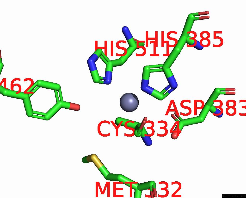
Mono view
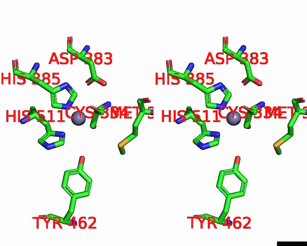
Stereo pair view
Mono view
Stereo pair view
A full contact list of Zinc with other atoms in the Zn binding
site number 1 of Crystal Structure of E. Coli Thrs Catalytic Domain Mutant G463S/Q484A within 5.0Å range:
|
Zinc binding site 2 out of 2 in 8wig
Go back to
Zinc binding site 2 out
of 2 in the Crystal Structure of E. Coli Thrs Catalytic Domain Mutant G463S/Q484A
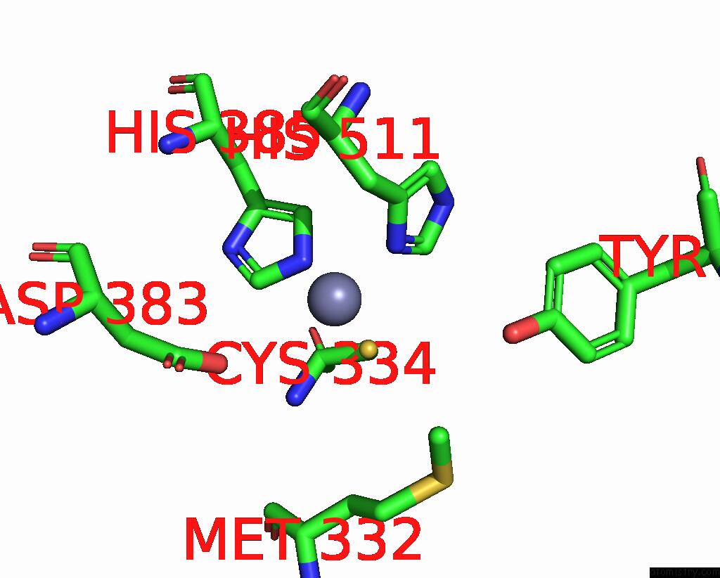
Mono view
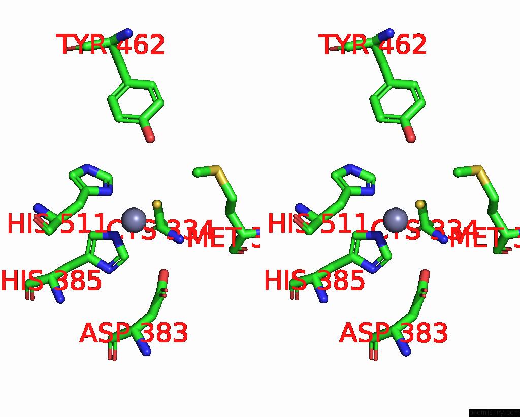
Stereo pair view
Mono view
Stereo pair view
A full contact list of Zinc with other atoms in the Zn binding
site number 2 of Crystal Structure of E. Coli Thrs Catalytic Domain Mutant G463S/Q484A within 5.0Å range:
|
Reference:
H.Qiao,
Z.Wang,
H.Yang,
M.Xia,
G.Yang,
F.Bai,
J.Wang,
P.Fang.
Specific Glycine-Dependent Enzyme Motion Determines the Potency of Conformation Selective Inhibitors of Threonyl-Trna Synthetase. Commun Biol V. 7 867 2024.
ISSN: ESSN 2399-3642
PubMed: 39014102
DOI: 10.1038/S42003-024-06559-X
Page generated: Fri Aug 22 15:12:33 2025
ISSN: ESSN 2399-3642
PubMed: 39014102
DOI: 10.1038/S42003-024-06559-X
Last articles
Zr in 1XC1Zr in 6Y7P
Zr in 6GNL
Zr in 6HYB
Zr in 4XYY
Zr in 5KHP
Zn in 9VXG
Zn in 9VWY
Zn in 9VCL
Zn in 9VKN