Zinc in PDB 8ufi: Cryo-Em Structure of Bovine Phosphodiesterase 6
Enzymatic activity of Cryo-Em Structure of Bovine Phosphodiesterase 6
All present enzymatic activity of Cryo-Em Structure of Bovine Phosphodiesterase 6:
3.1.4.35;
3.1.4.35;
Other elements in 8ufi:
The structure of Cryo-Em Structure of Bovine Phosphodiesterase 6 also contains other interesting chemical elements:
| Magnesium | (Mg) | 2 atoms |
Zinc Binding Sites:
The binding sites of Zinc atom in the Cryo-Em Structure of Bovine Phosphodiesterase 6
(pdb code 8ufi). This binding sites where shown within
5.0 Angstroms radius around Zinc atom.
In total 2 binding sites of Zinc where determined in the Cryo-Em Structure of Bovine Phosphodiesterase 6, PDB code: 8ufi:
Jump to Zinc binding site number: 1; 2;
In total 2 binding sites of Zinc where determined in the Cryo-Em Structure of Bovine Phosphodiesterase 6, PDB code: 8ufi:
Jump to Zinc binding site number: 1; 2;
Zinc binding site 1 out of 2 in 8ufi
Go back to
Zinc binding site 1 out
of 2 in the Cryo-Em Structure of Bovine Phosphodiesterase 6
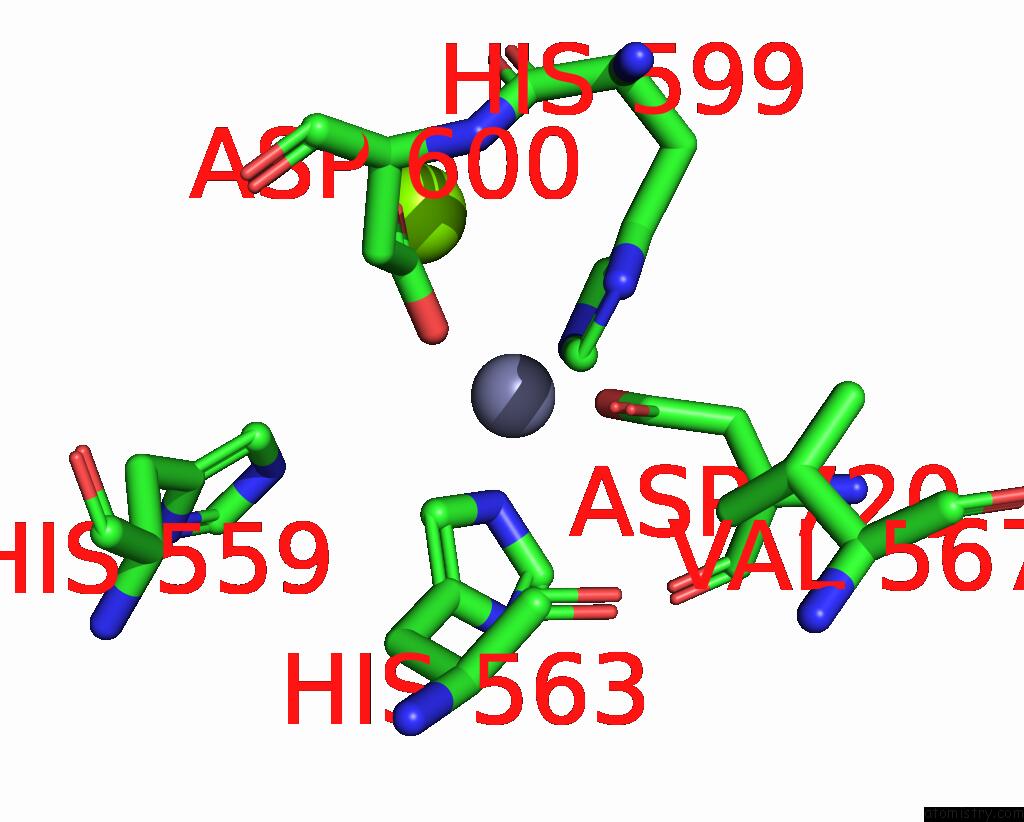
Mono view
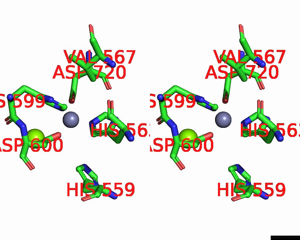
Stereo pair view
Mono view
Stereo pair view
A full contact list of Zinc with other atoms in the Zn binding
site number 1 of Cryo-Em Structure of Bovine Phosphodiesterase 6 within 5.0Å range:
|
Zinc binding site 2 out of 2 in 8ufi
Go back to
Zinc binding site 2 out
of 2 in the Cryo-Em Structure of Bovine Phosphodiesterase 6
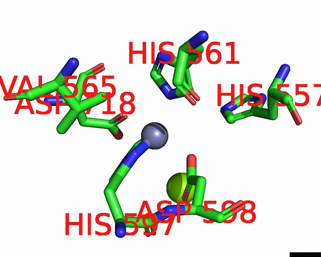
Mono view
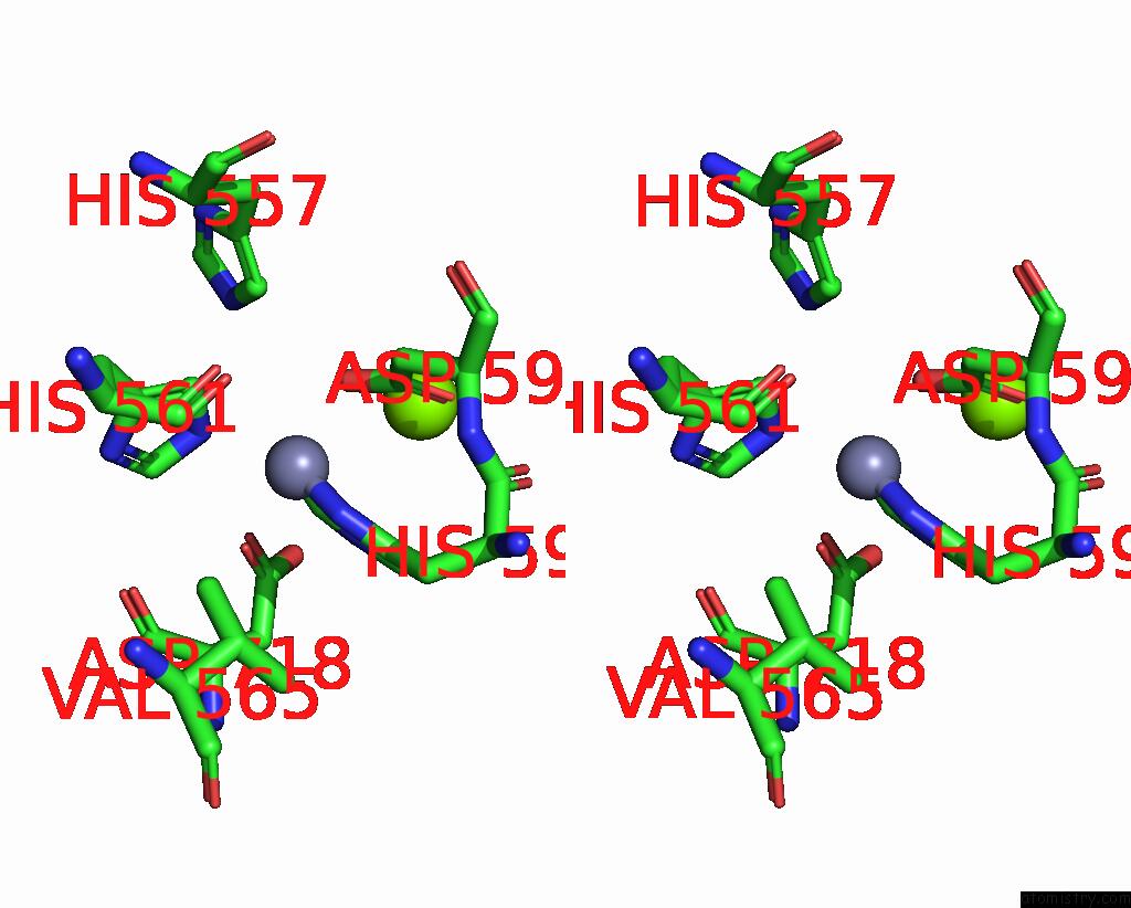
Stereo pair view
Mono view
Stereo pair view
A full contact list of Zinc with other atoms in the Zn binding
site number 2 of Cryo-Em Structure of Bovine Phosphodiesterase 6 within 5.0Å range:
|
Reference:
C.Aplin,
R.A.Cerione.
Probing the Mechanism By Which the Retinal G Protein Transducin Activates Its Biological Effector PDE6. J.Biol.Chem. 05608 2023.
ISSN: ESSN 1083-351X
PubMed: 38159849
DOI: 10.1016/J.JBC.2023.105608
Page generated: Thu Oct 31 12:07:08 2024
ISSN: ESSN 1083-351X
PubMed: 38159849
DOI: 10.1016/J.JBC.2023.105608
Last articles
Zn in 9MJ5Zn in 9HNW
Zn in 9G0L
Zn in 9FNE
Zn in 9DZN
Zn in 9E0I
Zn in 9D32
Zn in 9DAK
Zn in 8ZXC
Zn in 8ZUF