Zinc »
PDB 8s93-8sko »
8se1 »
Zinc in PDB 8se1: Structure of Full-Length Human Protein Kinase C Beta 2 (Pkcbii) in the Inactive Conformation
Enzymatic activity of Structure of Full-Length Human Protein Kinase C Beta 2 (Pkcbii) in the Inactive Conformation
All present enzymatic activity of Structure of Full-Length Human Protein Kinase C Beta 2 (Pkcbii) in the Inactive Conformation:
2.7.11.13;
2.7.11.13;
Protein crystallography data
The structure of Structure of Full-Length Human Protein Kinase C Beta 2 (Pkcbii) in the Inactive Conformation, PDB code: 8se1
was solved by
A.T.Q.Cong,
T.L.Witter,
E.S.Bruinsma,
S.Jayaraman,
M.B.Dugan,
J.R.Hawse,
M.P.Goetz,
M.J.Schellenberg,
with X-Ray Crystallography technique. A brief refinement statistics is given in the table below:
| Resolution Low / High (Å) | 48.75 / 3.32 |
| Space group | C 1 2 1 |
| Cell size a, b, c (Å), α, β, γ (°) | 130.556, 89.098, 160.714, 90, 107.05, 90 |
| R / Rfree (%) | 19.9 / 25.6 |
Other elements in 8se1:
The structure of Structure of Full-Length Human Protein Kinase C Beta 2 (Pkcbii) in the Inactive Conformation also contains other interesting chemical elements:
| Magnesium | (Mg) | 6 atoms |
Zinc Binding Sites:
The binding sites of Zinc atom in the Structure of Full-Length Human Protein Kinase C Beta 2 (Pkcbii) in the Inactive Conformation
(pdb code 8se1). This binding sites where shown within
5.0 Angstroms radius around Zinc atom.
In total 10 binding sites of Zinc where determined in the Structure of Full-Length Human Protein Kinase C Beta 2 (Pkcbii) in the Inactive Conformation, PDB code: 8se1:
Jump to Zinc binding site number: 1; 2; 3; 4; 5; 6; 7; 8; 9; 10;
In total 10 binding sites of Zinc where determined in the Structure of Full-Length Human Protein Kinase C Beta 2 (Pkcbii) in the Inactive Conformation, PDB code: 8se1:
Jump to Zinc binding site number: 1; 2; 3; 4; 5; 6; 7; 8; 9; 10;
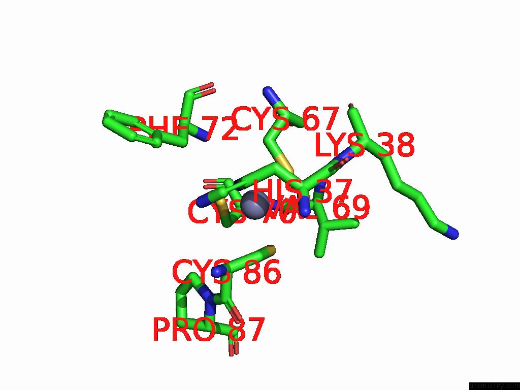
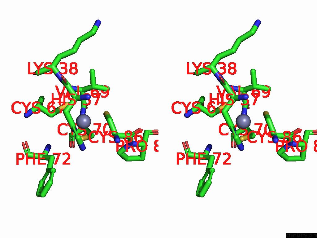
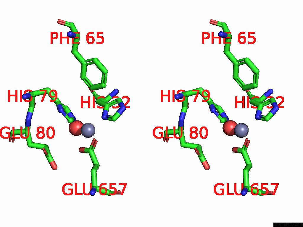
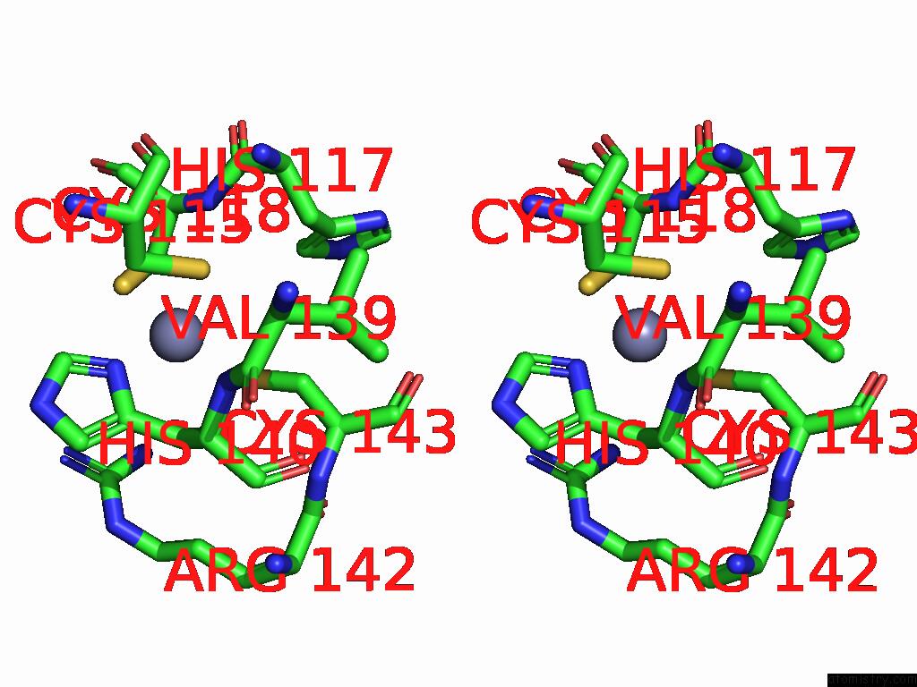
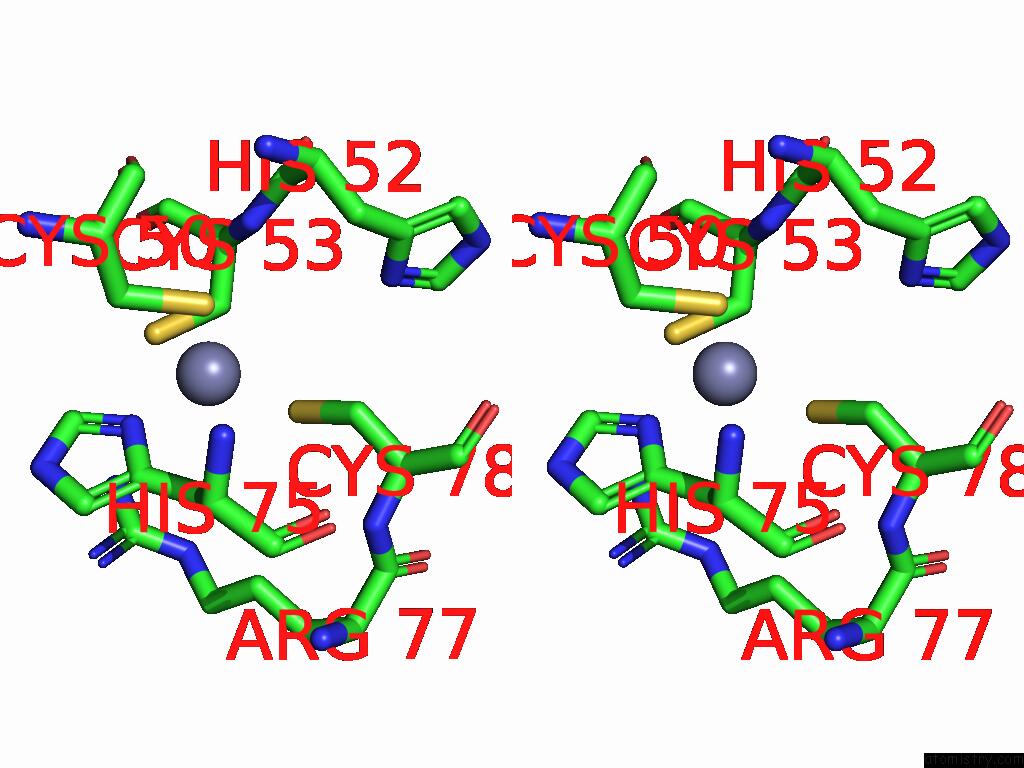
Zinc binding site 1 out of 10 in 8se1
Go back to
Zinc binding site 1 out
of 10 in the Structure of Full-Length Human Protein Kinase C Beta 2 (Pkcbii) in the Inactive Conformation
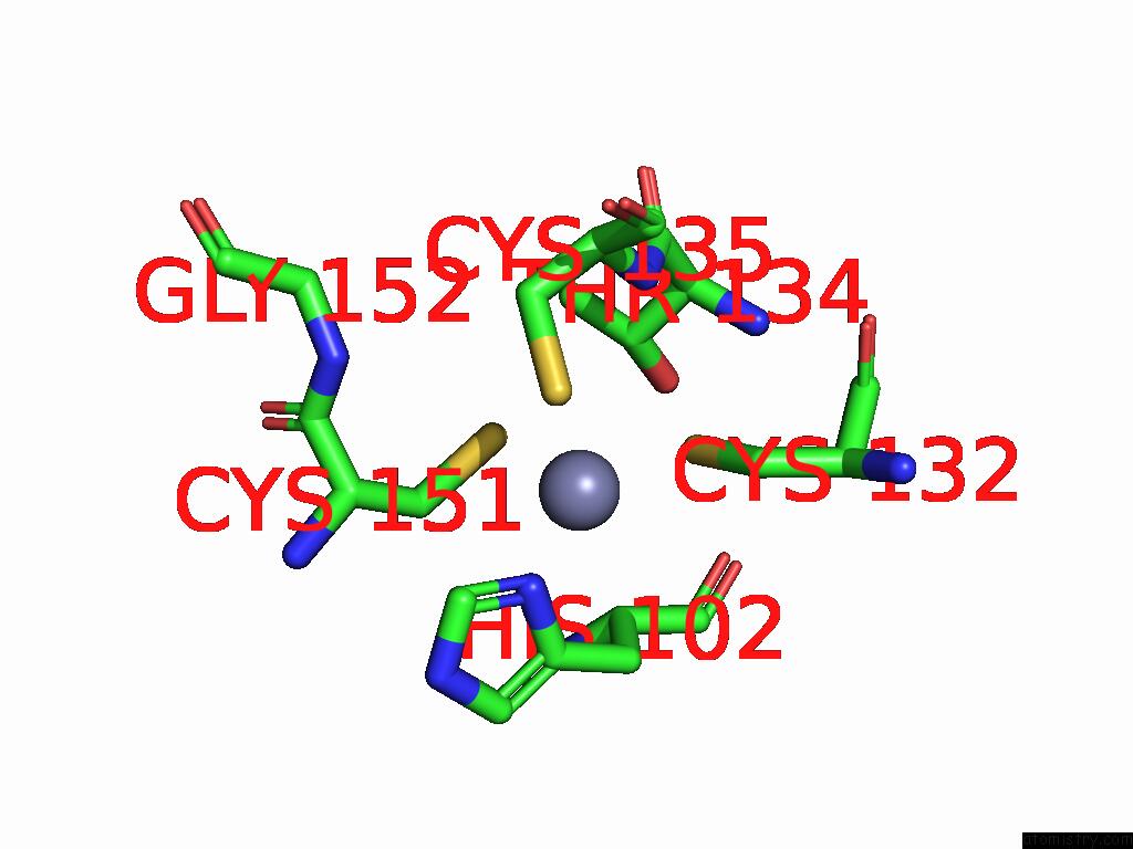
Mono view
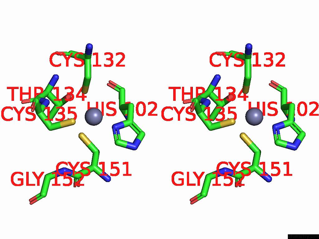
Stereo pair view
Mono view
Stereo pair view
A full contact list of Zinc with other atoms in the Zn binding
site number 1 of Structure of Full-Length Human Protein Kinase C Beta 2 (Pkcbii) in the Inactive Conformation within 5.0Å range:
|
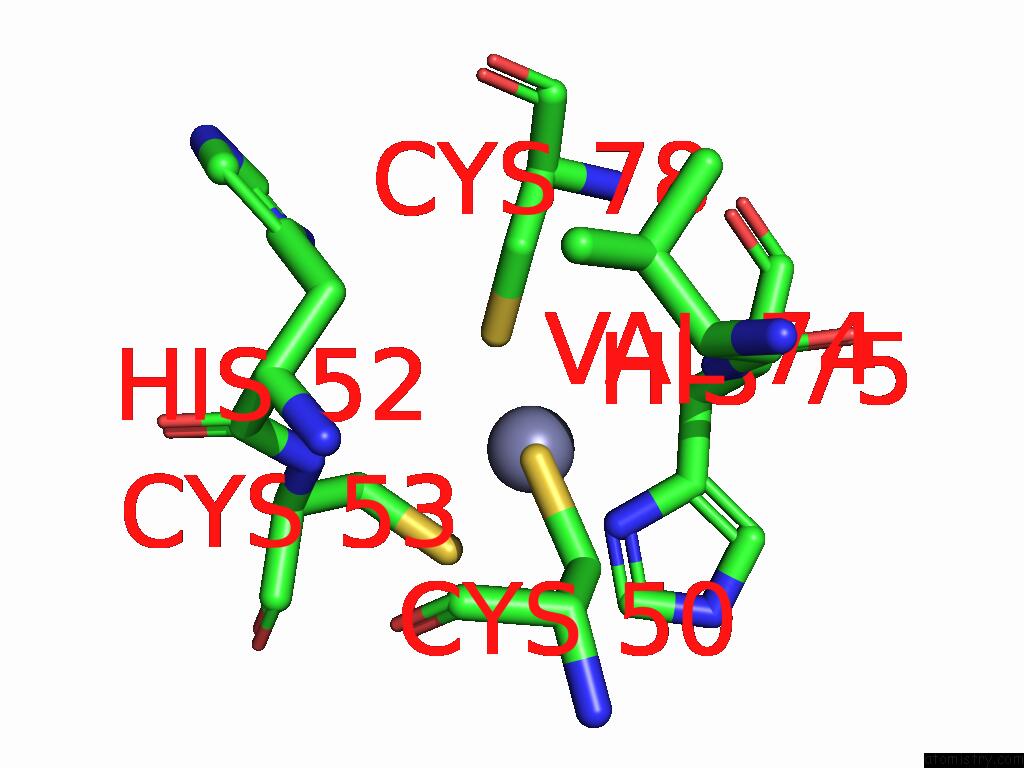
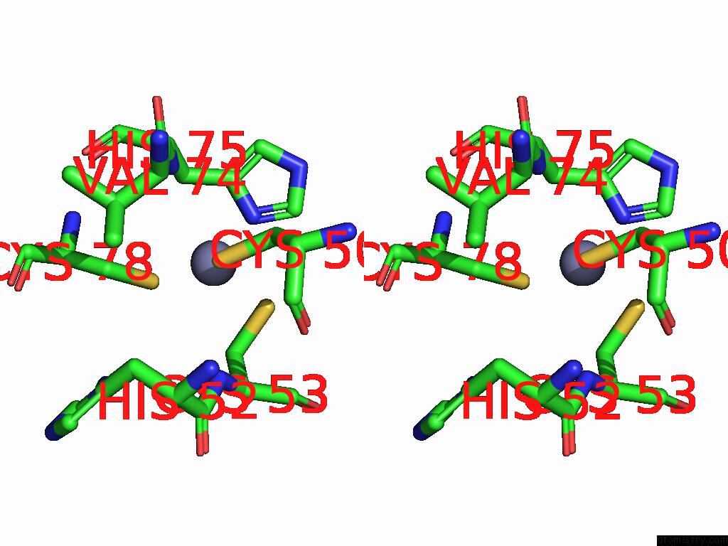
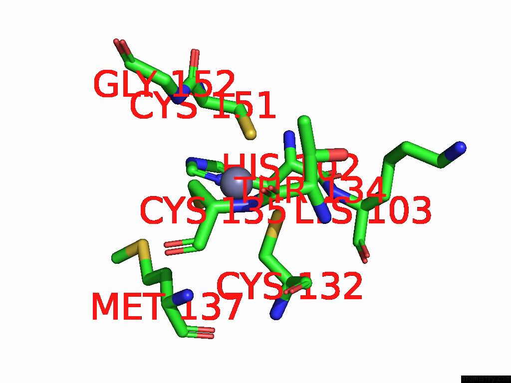
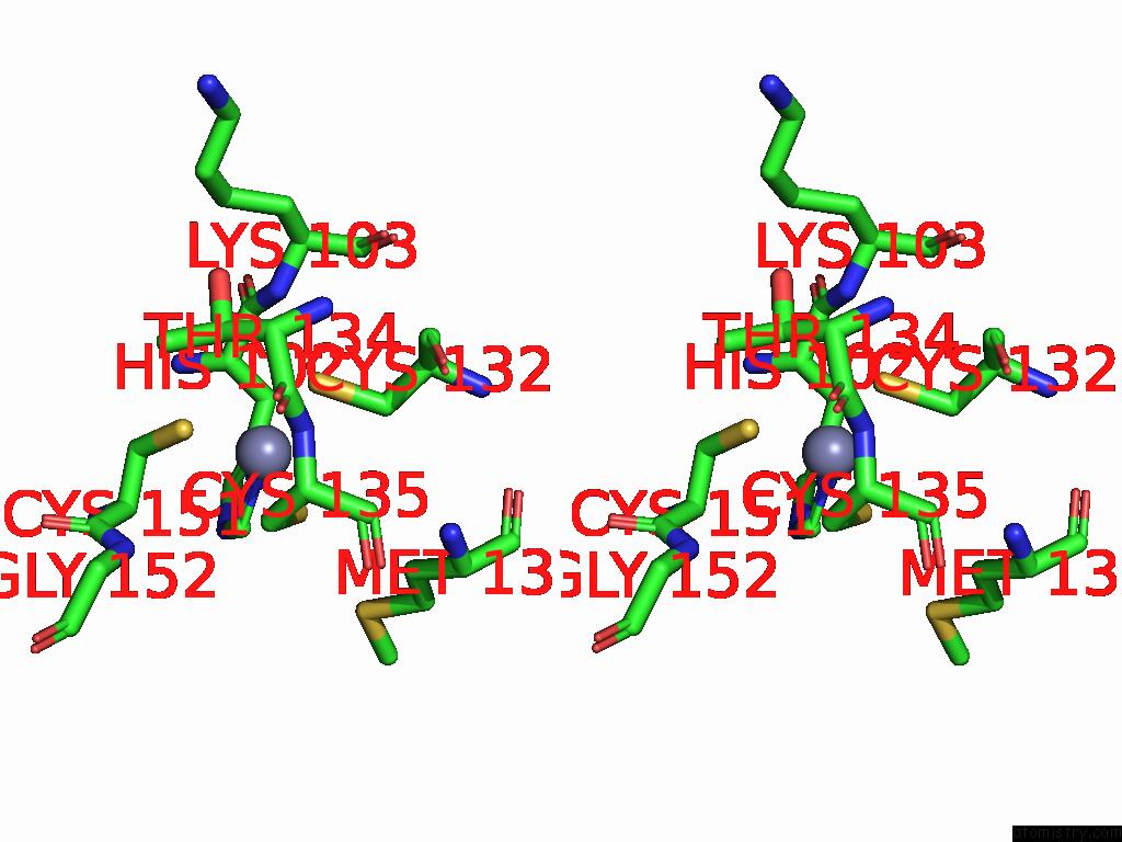
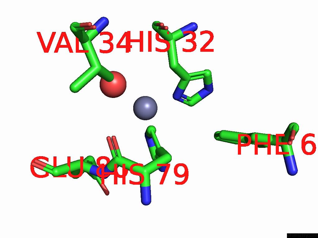
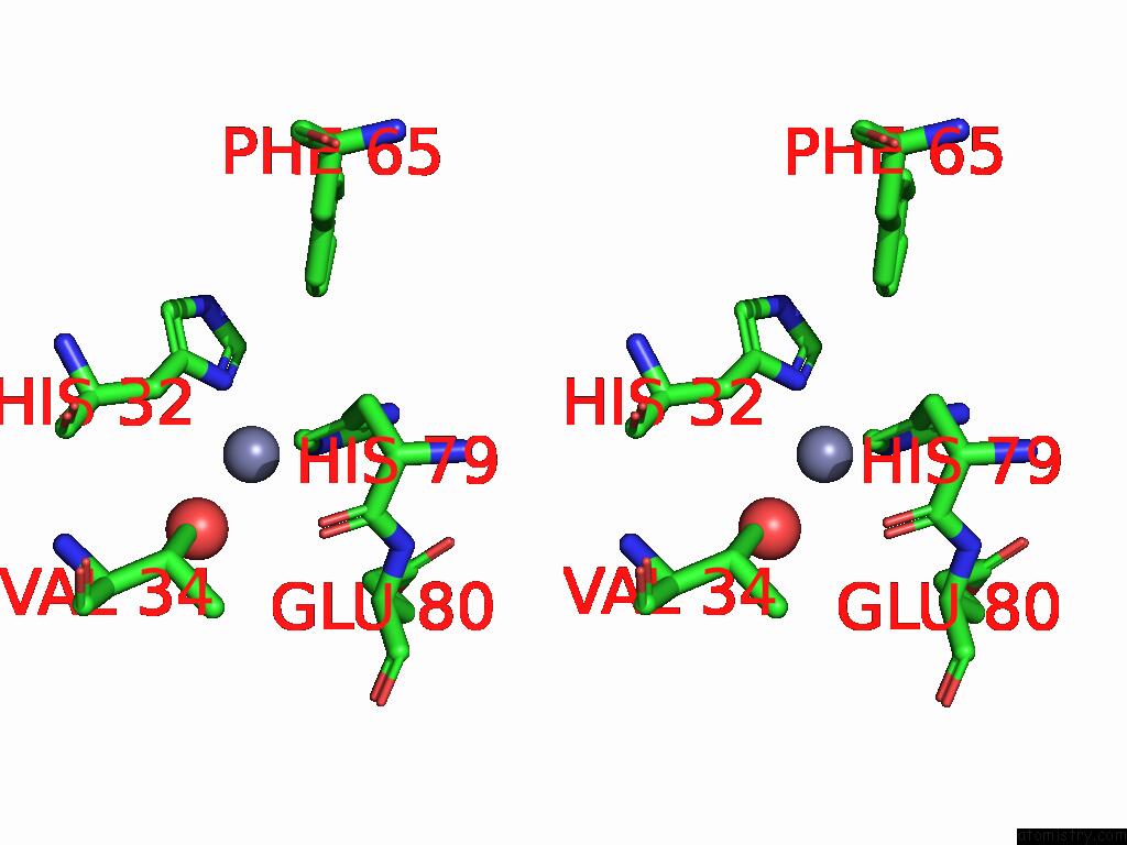
Zinc binding site 2 out of 10 in 8se1
Go back to
Zinc binding site 2 out
of 10 in the Structure of Full-Length Human Protein Kinase C Beta 2 (Pkcbii) in the Inactive Conformation
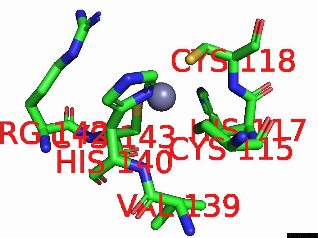
Mono view
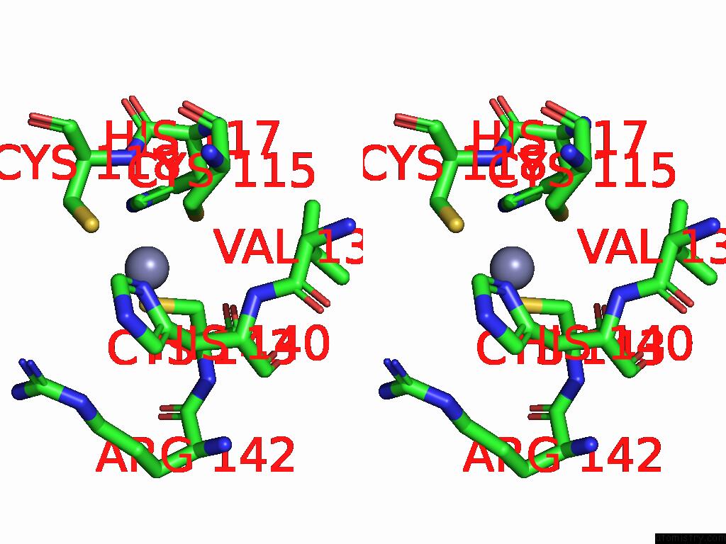
Stereo pair view
Mono view
Stereo pair view
A full contact list of Zinc with other atoms in the Zn binding
site number 2 of Structure of Full-Length Human Protein Kinase C Beta 2 (Pkcbii) in the Inactive Conformation within 5.0Å range:
|
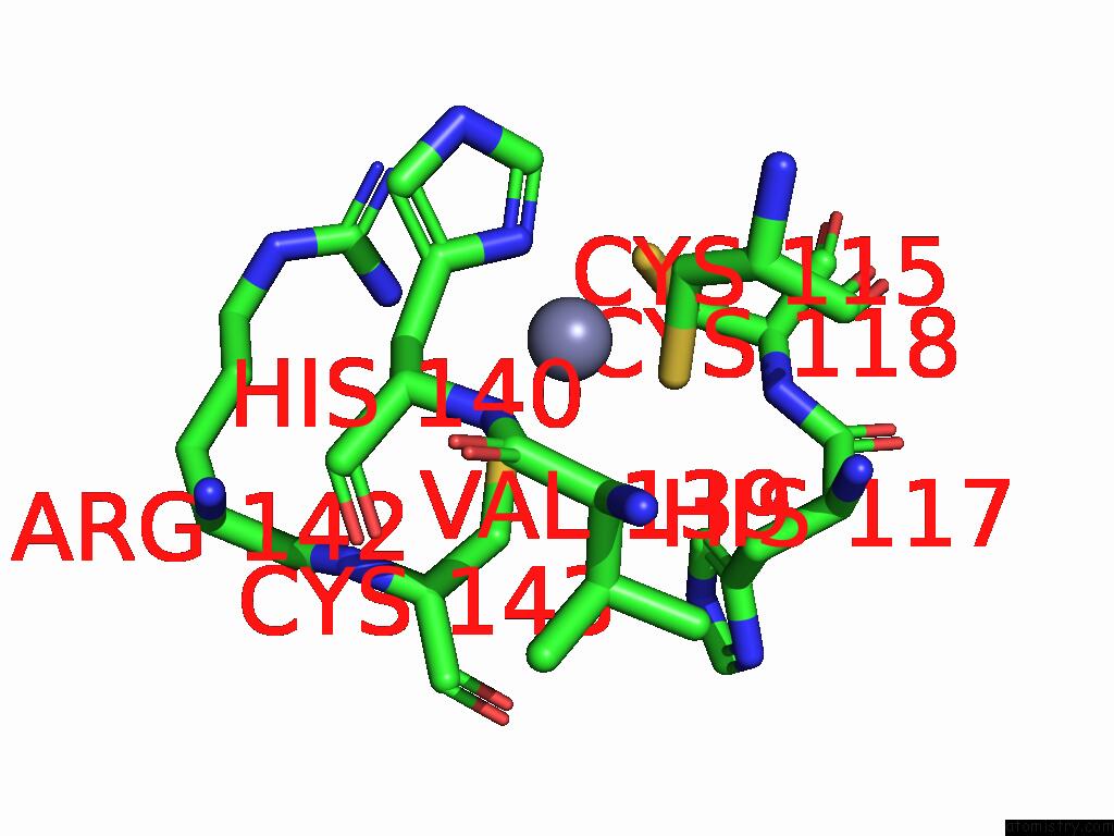
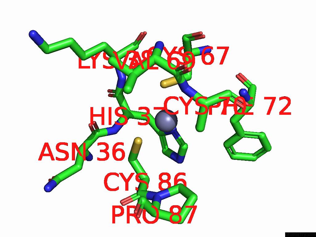
Zinc binding site 3 out of 10 in 8se1
Go back to
Zinc binding site 3 out
of 10 in the Structure of Full-Length Human Protein Kinase C Beta 2 (Pkcbii) in the Inactive Conformation
Mono view
Stereo pair view
Mono view
Stereo pair view
A full contact list of Zinc with other atoms in the Zn binding
site number 3 of Structure of Full-Length Human Protein Kinase C Beta 2 (Pkcbii) in the Inactive Conformation within 5.0Å range:
|
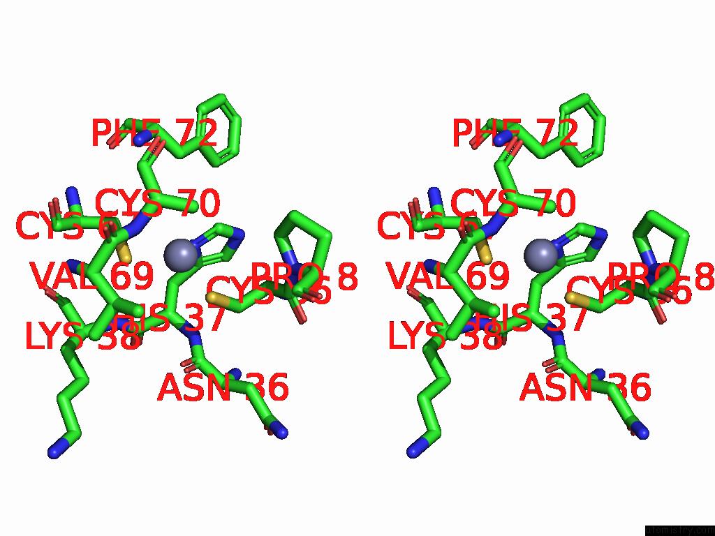
Zinc binding site 4 out of 10 in 8se1
Go back to
Zinc binding site 4 out
of 10 in the Structure of Full-Length Human Protein Kinase C Beta 2 (Pkcbii) in the Inactive Conformation
Mono view
Stereo pair view
Mono view
Stereo pair view
A full contact list of Zinc with other atoms in the Zn binding
site number 4 of Structure of Full-Length Human Protein Kinase C Beta 2 (Pkcbii) in the Inactive Conformation within 5.0Å range:
|
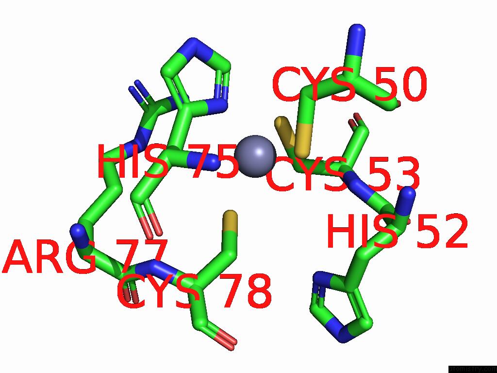
Zinc binding site 5 out of 10 in 8se1
Go back to
Zinc binding site 5 out
of 10 in the Structure of Full-Length Human Protein Kinase C Beta 2 (Pkcbii) in the Inactive Conformation

Mono view
Stereo pair view
Mono view
Stereo pair view
A full contact list of Zinc with other atoms in the Zn binding
site number 5 of Structure of Full-Length Human Protein Kinase C Beta 2 (Pkcbii) in the Inactive Conformation within 5.0Å range:
|
Zinc binding site 6 out of 10 in 8se1
Go back to
Zinc binding site 6 out
of 10 in the Structure of Full-Length Human Protein Kinase C Beta 2 (Pkcbii) in the Inactive Conformation
Mono view
Stereo pair view
Mono view
Stereo pair view
A full contact list of Zinc with other atoms in the Zn binding
site number 6 of Structure of Full-Length Human Protein Kinase C Beta 2 (Pkcbii) in the Inactive Conformation within 5.0Å range:
|
Zinc binding site 7 out of 10 in 8se1
Go back to
Zinc binding site 7 out
of 10 in the Structure of Full-Length Human Protein Kinase C Beta 2 (Pkcbii) in the Inactive Conformation
Mono view
Stereo pair view
Mono view
Stereo pair view
A full contact list of Zinc with other atoms in the Zn binding
site number 7 of Structure of Full-Length Human Protein Kinase C Beta 2 (Pkcbii) in the Inactive Conformation within 5.0Å range:
|
Zinc binding site 8 out of 10 in 8se1
Go back to
Zinc binding site 8 out
of 10 in the Structure of Full-Length Human Protein Kinase C Beta 2 (Pkcbii) in the Inactive Conformation
Mono view
Stereo pair view
Mono view
Stereo pair view
A full contact list of Zinc with other atoms in the Zn binding
site number 8 of Structure of Full-Length Human Protein Kinase C Beta 2 (Pkcbii) in the Inactive Conformation within 5.0Å range:
|
Zinc binding site 9 out of 10 in 8se1
Go back to
Zinc binding site 9 out
of 10 in the Structure of Full-Length Human Protein Kinase C Beta 2 (Pkcbii) in the Inactive Conformation
Mono view
Stereo pair view
Mono view
Stereo pair view
A full contact list of Zinc with other atoms in the Zn binding
site number 9 of Structure of Full-Length Human Protein Kinase C Beta 2 (Pkcbii) in the Inactive Conformation within 5.0Å range:
|
Zinc binding site 10 out of 10 in 8se1
Go back to
Zinc binding site 10 out
of 10 in the Structure of Full-Length Human Protein Kinase C Beta 2 (Pkcbii) in the Inactive Conformation
Mono view
Stereo pair view
Mono view
Stereo pair view
A full contact list of Zinc with other atoms in the Zn binding
site number 10 of Structure of Full-Length Human Protein Kinase C Beta 2 (Pkcbii) in the Inactive Conformation within 5.0Å range:
|
Reference:
A.T.Q.Cong,
T.L.Witter,
E.S.Bruinsma,
S.S.Bhattacharya,
S.Jayaraman,
M.B.Dugan,
J.Paluncic,
M.J.Kuffel,
J.Farmakes,
J.Alvey,
X.Wu,
A.P.Fields,
A.Pandey,
J.R.Hawse,
M.P.Goetz,
M.J.Schellenberg.
Molecular Basis of Allosteric Regulation and Pharmaceutical Targeting of Protein Kinase C B To Be Published.
Page generated: Tue Aug 26 21:59:00 2025
Last articles
Zn in 9QM9Zn in 9S44
Zn in 9OFE
Zn in 9OFC
Zn in 9OFD
Zn in 9OF1
Zn in 9OFB
Zn in 9N0J
Zn in 9M5X
Zn in 9LGI