Zinc in PDB 8jpw: Crystal Structure of Single-Chain L-Glutamate Oxidase Mutant From Streptomyces Sp. X-119-6
Protein crystallography data
The structure of Crystal Structure of Single-Chain L-Glutamate Oxidase Mutant From Streptomyces Sp. X-119-6, PDB code: 8jpw
was solved by
H.Yamaguchi,
K.Takahashi,
M.Tatsumi,
U.Tagami,
T.Mizukoshi,
H.Miyano,
M.Sugiki,
with X-Ray Crystallography technique. A brief refinement statistics is given in the table below:
| Resolution Low / High (Å) | 49.32 / 2.66 |
| Space group | I 4 2 2 |
| Cell size a, b, c (Å), α, β, γ (°) | 158.4, 158.4, 138.235, 90, 90, 90 |
| R / Rfree (%) | 25.9 / 30.5 |
Zinc Binding Sites:
The binding sites of Zinc atom in the Crystal Structure of Single-Chain L-Glutamate Oxidase Mutant From Streptomyces Sp. X-119-6
(pdb code 8jpw). This binding sites where shown within
5.0 Angstroms radius around Zinc atom.
In total 2 binding sites of Zinc where determined in the Crystal Structure of Single-Chain L-Glutamate Oxidase Mutant From Streptomyces Sp. X-119-6, PDB code: 8jpw:
Jump to Zinc binding site number: 1; 2;
In total 2 binding sites of Zinc where determined in the Crystal Structure of Single-Chain L-Glutamate Oxidase Mutant From Streptomyces Sp. X-119-6, PDB code: 8jpw:
Jump to Zinc binding site number: 1; 2;
Zinc binding site 1 out of 2 in 8jpw
Go back to
Zinc binding site 1 out
of 2 in the Crystal Structure of Single-Chain L-Glutamate Oxidase Mutant From Streptomyces Sp. X-119-6
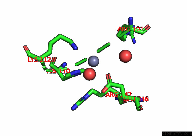
Mono view
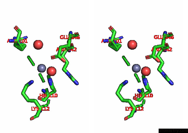
Stereo pair view
Mono view
Stereo pair view
A full contact list of Zinc with other atoms in the Zn binding
site number 1 of Crystal Structure of Single-Chain L-Glutamate Oxidase Mutant From Streptomyces Sp. X-119-6 within 5.0Å range:
|
Zinc binding site 2 out of 2 in 8jpw
Go back to
Zinc binding site 2 out
of 2 in the Crystal Structure of Single-Chain L-Glutamate Oxidase Mutant From Streptomyces Sp. X-119-6
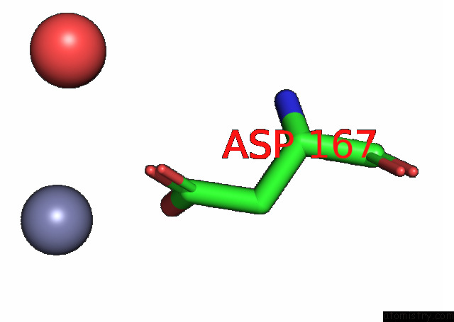
Mono view
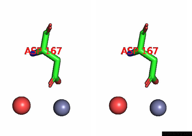
Stereo pair view
Mono view
Stereo pair view
A full contact list of Zinc with other atoms in the Zn binding
site number 2 of Crystal Structure of Single-Chain L-Glutamate Oxidase Mutant From Streptomyces Sp. X-119-6 within 5.0Å range:
|
Reference:
H.Yamaguchi,
K.Takahashi,
M.Tatsumi,
U.Tagami,
T.Mizukoshi,
H.Miyano,
M.Sugiki.
Development of A Novel Single-Chain L-Glutamate Oxidase From Streptomyces Sp. X-119-6 By Inserting Flexible Linkers Enzyme.Microb.Technol. V. 170 10287 2023.
ISSN: ISSN 0141-0229
DOI: 10.1016/J.ENZMICTEC.2023.110287
Page generated: Thu Oct 31 08:24:12 2024
ISSN: ISSN 0141-0229
DOI: 10.1016/J.ENZMICTEC.2023.110287
Last articles
Zn in 9MJ5Zn in 9HNW
Zn in 9G0L
Zn in 9FNE
Zn in 9DZN
Zn in 9E0I
Zn in 9D32
Zn in 9DAK
Zn in 8ZXC
Zn in 8ZUF