Zinc in PDB 8j2p: Crystal Structure of Pml B-BOX2
Protein crystallography data
The structure of Crystal Structure of Pml B-BOX2, PDB code: 8j2p
was solved by
C.Zhou,
N.Zang,
J.Zhang,
with X-Ray Crystallography technique. A brief refinement statistics is given in the table below:
| Resolution Low / High (Å) | 35.38 / 2.09 |
| Space group | H 3 |
| Cell size a, b, c (Å), α, β, γ (°) | 122.55, 122.55, 163.224, 90, 90, 120 |
| R / Rfree (%) | 19.1 / 23.5 |
Zinc Binding Sites:
The binding sites of Zinc atom in the Crystal Structure of Pml B-BOX2
(pdb code 8j2p). This binding sites where shown within
5.0 Angstroms radius around Zinc atom.
In total 4 binding sites of Zinc where determined in the Crystal Structure of Pml B-BOX2, PDB code: 8j2p:
Jump to Zinc binding site number: 1; 2; 3; 4;
In total 4 binding sites of Zinc where determined in the Crystal Structure of Pml B-BOX2, PDB code: 8j2p:
Jump to Zinc binding site number: 1; 2; 3; 4;
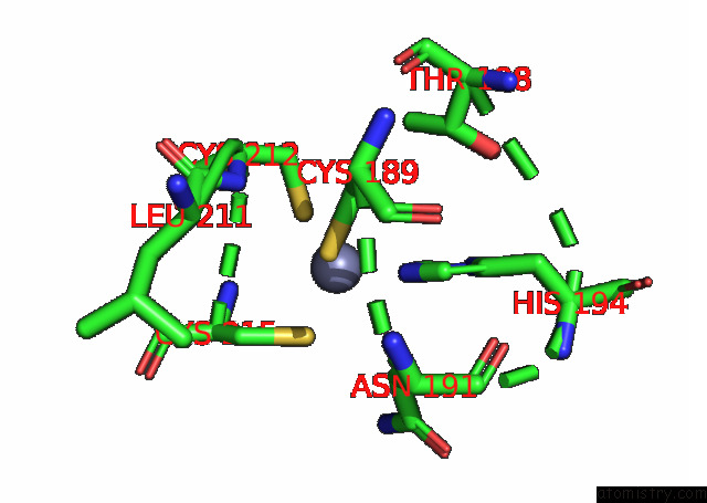
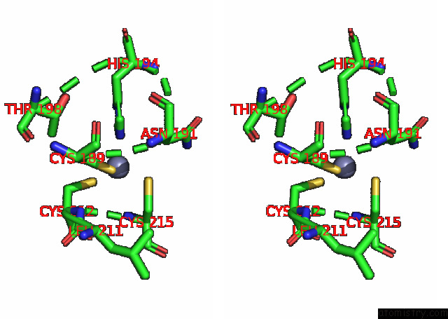
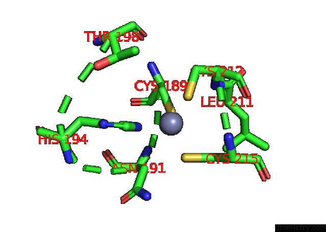
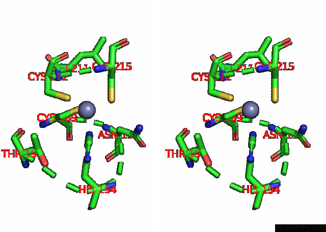
Zinc binding site 1 out of 4 in 8j2p
Go back to
Zinc binding site 1 out
of 4 in the Crystal Structure of Pml B-BOX2
Mono view
Stereo pair view
Mono view
Stereo pair view
A full contact list of Zinc with other atoms in the Zn binding
site number 1 of Crystal Structure of Pml B-BOX2 within 5.0Å range:
|
Zinc binding site 2 out of 4 in 8j2p
Go back to
Zinc binding site 2 out
of 4 in the Crystal Structure of Pml B-BOX2
Mono view
Stereo pair view
Mono view
Stereo pair view
A full contact list of Zinc with other atoms in the Zn binding
site number 2 of Crystal Structure of Pml B-BOX2 within 5.0Å range:
|
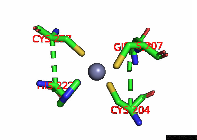
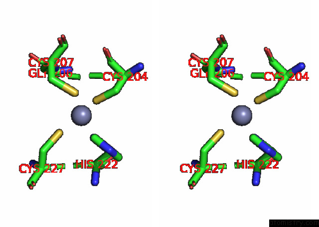
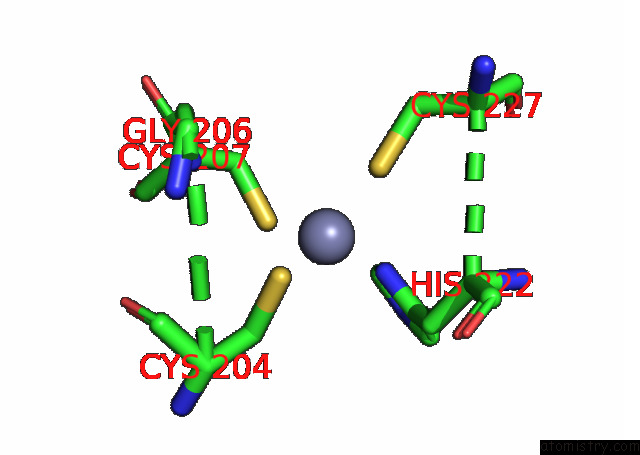
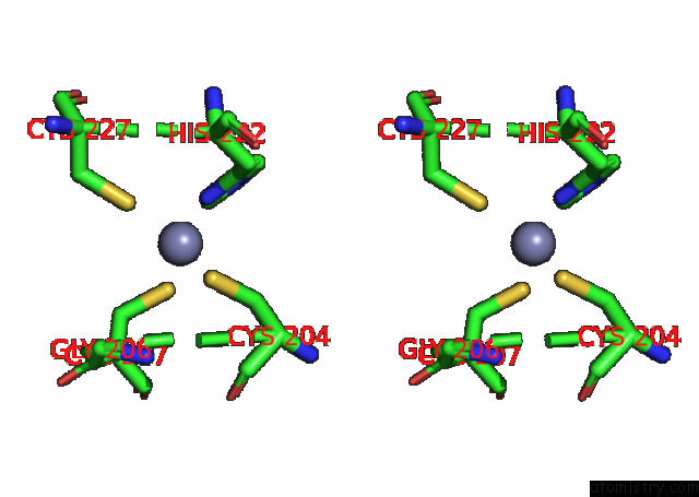
Zinc binding site 3 out of 4 in 8j2p
Go back to
Zinc binding site 3 out
of 4 in the Crystal Structure of Pml B-BOX2
Mono view
Stereo pair view
Mono view
Stereo pair view
A full contact list of Zinc with other atoms in the Zn binding
site number 3 of Crystal Structure of Pml B-BOX2 within 5.0Å range:
|
Zinc binding site 4 out of 4 in 8j2p
Go back to
Zinc binding site 4 out
of 4 in the Crystal Structure of Pml B-BOX2
Mono view
Stereo pair view
Mono view
Stereo pair view
A full contact list of Zinc with other atoms in the Zn binding
site number 4 of Crystal Structure of Pml B-BOX2 within 5.0Å range:
|
Reference:
P.Bercier,
Q.Q.Wang,
N.Zang,
J.Zhang,
C.Yang,
Y.Maimaitiyiming,
M.Abou-Ghali,
C.Berthier,
C.Wu,
M.Niwa-Kawakita,
T.Dirami,
M.C.Geoffroy,
O.Ferhi,
S.Quentin,
S.Benhenda,
Y.Ogra,
Z.Gueroui,
C.Zhou,
H.Naranmandura,
H.De The,
V.Lallemand-Breitenbach.
Structural Basis of Pml/Rara Oncoprotein Targeting By Arsenic Unravels A Cysteine Rheostat Controlling Pml Body Assembly and Function. Cancer Discov 2023.
ISSN: ESSN 2159-8290
PubMed: 37655965
DOI: 10.1158/2159-8290.CD-23-0453
Page generated: Thu Oct 31 08:03:26 2024
ISSN: ESSN 2159-8290
PubMed: 37655965
DOI: 10.1158/2159-8290.CD-23-0453
Last articles
Zn in 9MJ5Zn in 9HNW
Zn in 9G0L
Zn in 9FNE
Zn in 9DZN
Zn in 9E0I
Zn in 9D32
Zn in 9DAK
Zn in 8ZXC
Zn in 8ZUF