Zinc in PDB 8imd: Crystal Structure of Cu/Zn Superoxide Dismutase From Paenibacillus Lautus
Protein crystallography data
The structure of Crystal Structure of Cu/Zn Superoxide Dismutase From Paenibacillus Lautus, PDB code: 8imd
was solved by
S.Narikiyo,
Y.Furukawa,
M.Akutsu,
with X-Ray Crystallography technique. A brief refinement statistics is given in the table below:
| Resolution Low / High (Å) | 45.35 / 1.45 |
| Space group | P 21 21 21 |
| Cell size a, b, c (Å), α, β, γ (°) | 45.24, 53.612, 169.567, 90, 90, 90 |
| R / Rfree (%) | 18.8 / 22.1 |
Other elements in 8imd:
The structure of Crystal Structure of Cu/Zn Superoxide Dismutase From Paenibacillus Lautus also contains other interesting chemical elements:
| Copper | (Cu) | 2 atoms |
Zinc Binding Sites:
The binding sites of Zinc atom in the Crystal Structure of Cu/Zn Superoxide Dismutase From Paenibacillus Lautus
(pdb code 8imd). This binding sites where shown within
5.0 Angstroms radius around Zinc atom.
In total 2 binding sites of Zinc where determined in the Crystal Structure of Cu/Zn Superoxide Dismutase From Paenibacillus Lautus, PDB code: 8imd:
Jump to Zinc binding site number: 1; 2;
In total 2 binding sites of Zinc where determined in the Crystal Structure of Cu/Zn Superoxide Dismutase From Paenibacillus Lautus, PDB code: 8imd:
Jump to Zinc binding site number: 1; 2;
Zinc binding site 1 out of 2 in 8imd
Go back to
Zinc binding site 1 out
of 2 in the Crystal Structure of Cu/Zn Superoxide Dismutase From Paenibacillus Lautus
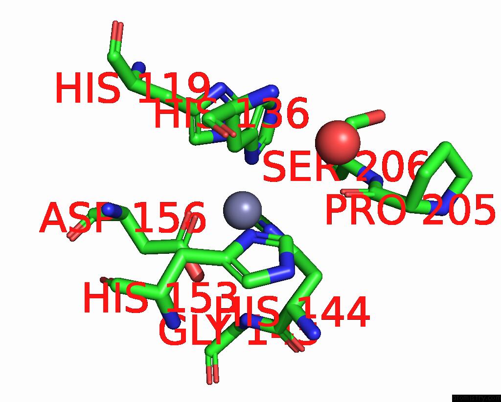
Mono view

Stereo pair view
Mono view
Stereo pair view
A full contact list of Zinc with other atoms in the Zn binding
site number 1 of Crystal Structure of Cu/Zn Superoxide Dismutase From Paenibacillus Lautus within 5.0Å range:
|
Zinc binding site 2 out of 2 in 8imd
Go back to
Zinc binding site 2 out
of 2 in the Crystal Structure of Cu/Zn Superoxide Dismutase From Paenibacillus Lautus
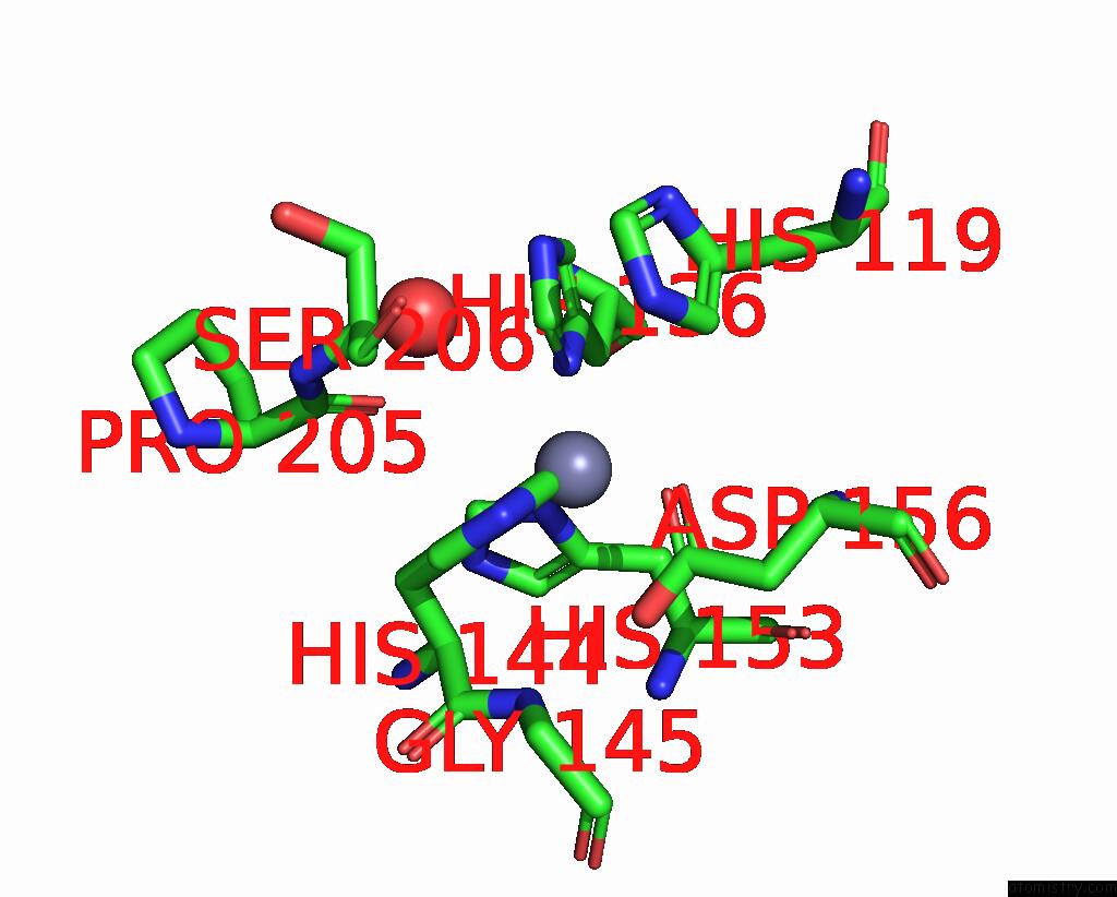
Mono view
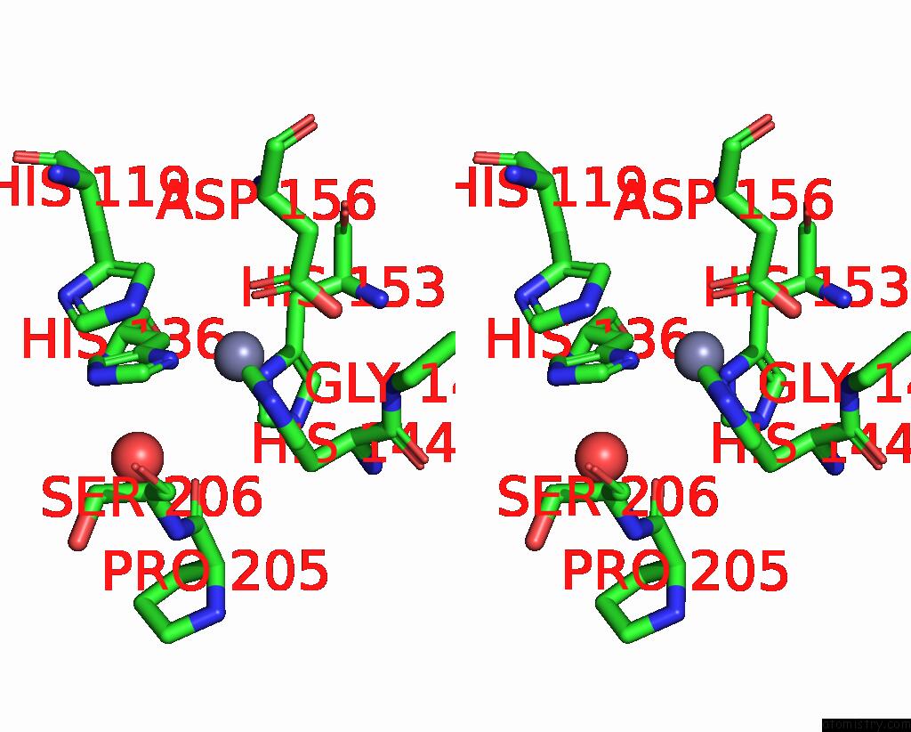
Stereo pair view
Mono view
Stereo pair view
A full contact list of Zinc with other atoms in the Zn binding
site number 2 of Crystal Structure of Cu/Zn Superoxide Dismutase From Paenibacillus Lautus within 5.0Å range:
|
Reference:
Y.Furukawa,
A.Shintani,
S.Narikiyo,
K.Sue,
M.Akutsu,
N.Muraki.
Characterization of A Novel Cysteine-Less Cu/Zn-Superoxide Dismutase in Paenibacillus Lautus Missing A Conserved Disulfide Bond. J.Biol.Chem. V. 299 05040 2023.
ISSN: ESSN 1083-351X
PubMed: 37442237
DOI: 10.1016/J.JBC.2023.105040
Page generated: Thu Oct 31 07:53:03 2024
ISSN: ESSN 1083-351X
PubMed: 37442237
DOI: 10.1016/J.JBC.2023.105040
Last articles
Zn in 9MJ5Zn in 9HNW
Zn in 9G0L
Zn in 9FNE
Zn in 9DZN
Zn in 9E0I
Zn in 9D32
Zn in 9DAK
Zn in 8ZXC
Zn in 8ZUF