Zinc in PDB 8hgz: Crystal Structure of Insulin
Protein crystallography data
The structure of Crystal Structure of Insulin, PDB code: 8hgz
was solved by
H.Demirci,
E.Ayan,
with X-Ray Crystallography technique. A brief refinement statistics is given in the table below:
| Resolution Low / High (Å) | 21.64 / 1.70 |
| Space group | H 3 |
| Cell size a, b, c (Å), α, β, γ (°) | 78.885, 78.885, 79.272, 90, 90, 120 |
| R / Rfree (%) | 21.8 / 25.8 |
Other elements in 8hgz:
The structure of Crystal Structure of Insulin also contains other interesting chemical elements:
| Chlorine | (Cl) | 4 atoms |
Zinc Binding Sites:
The binding sites of Zinc atom in the Crystal Structure of Insulin
(pdb code 8hgz). This binding sites where shown within
5.0 Angstroms radius around Zinc atom.
In total 4 binding sites of Zinc where determined in the Crystal Structure of Insulin, PDB code: 8hgz:
Jump to Zinc binding site number: 1; 2; 3; 4;
In total 4 binding sites of Zinc where determined in the Crystal Structure of Insulin, PDB code: 8hgz:
Jump to Zinc binding site number: 1; 2; 3; 4;
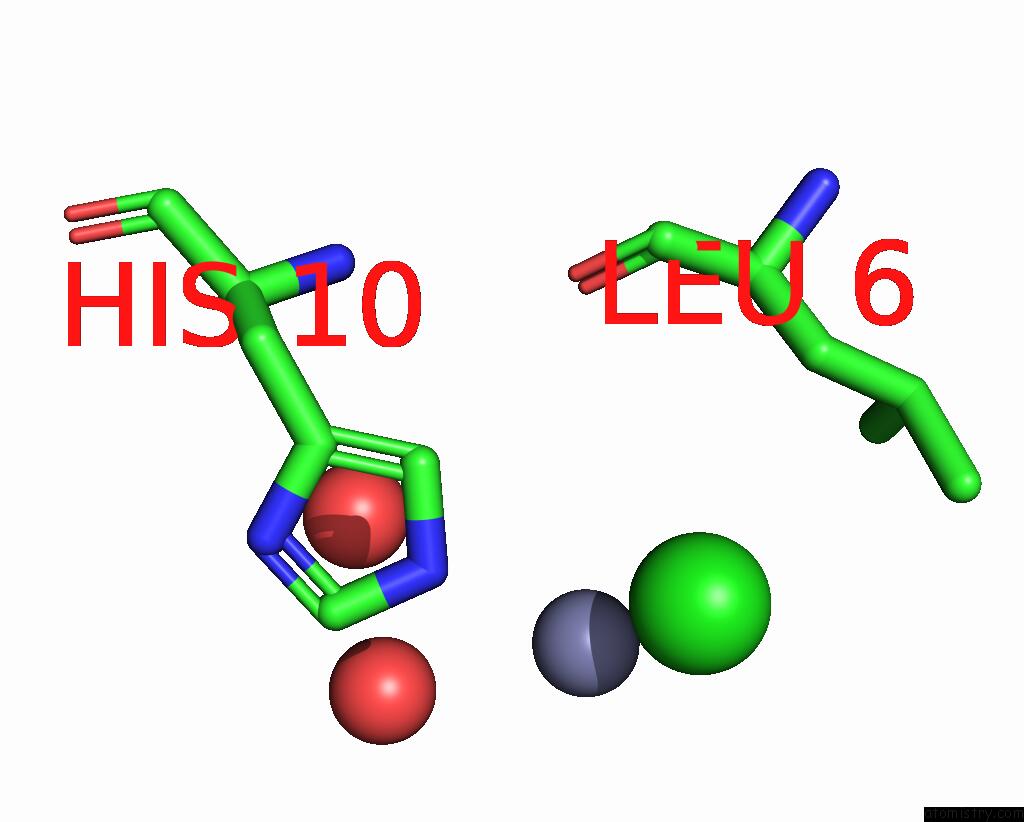
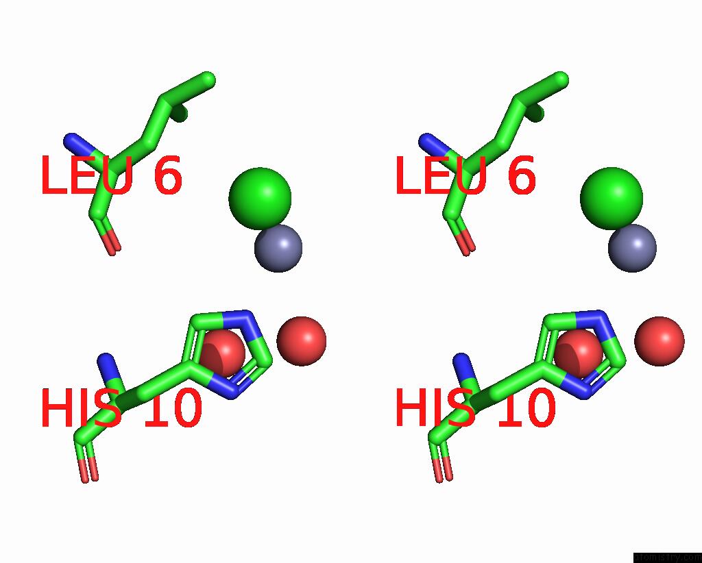
Zinc binding site 1 out of 4 in 8hgz
Go back to
Zinc binding site 1 out
of 4 in the Crystal Structure of Insulin
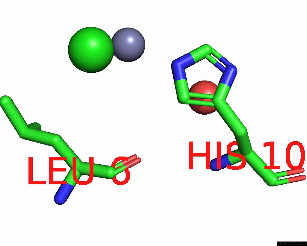
Mono view
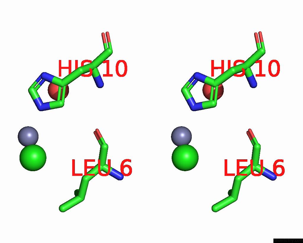
Stereo pair view
Mono view
Stereo pair view
A full contact list of Zinc with other atoms in the Zn binding
site number 1 of Crystal Structure of Insulin within 5.0Å range:
|
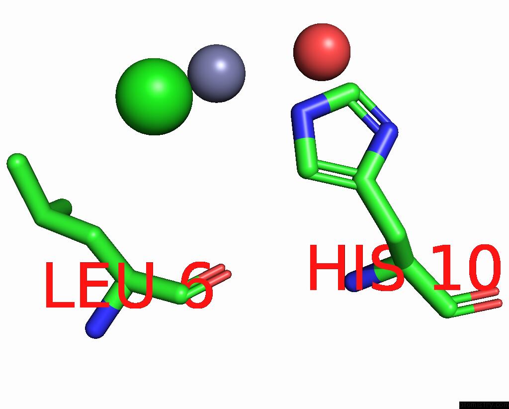
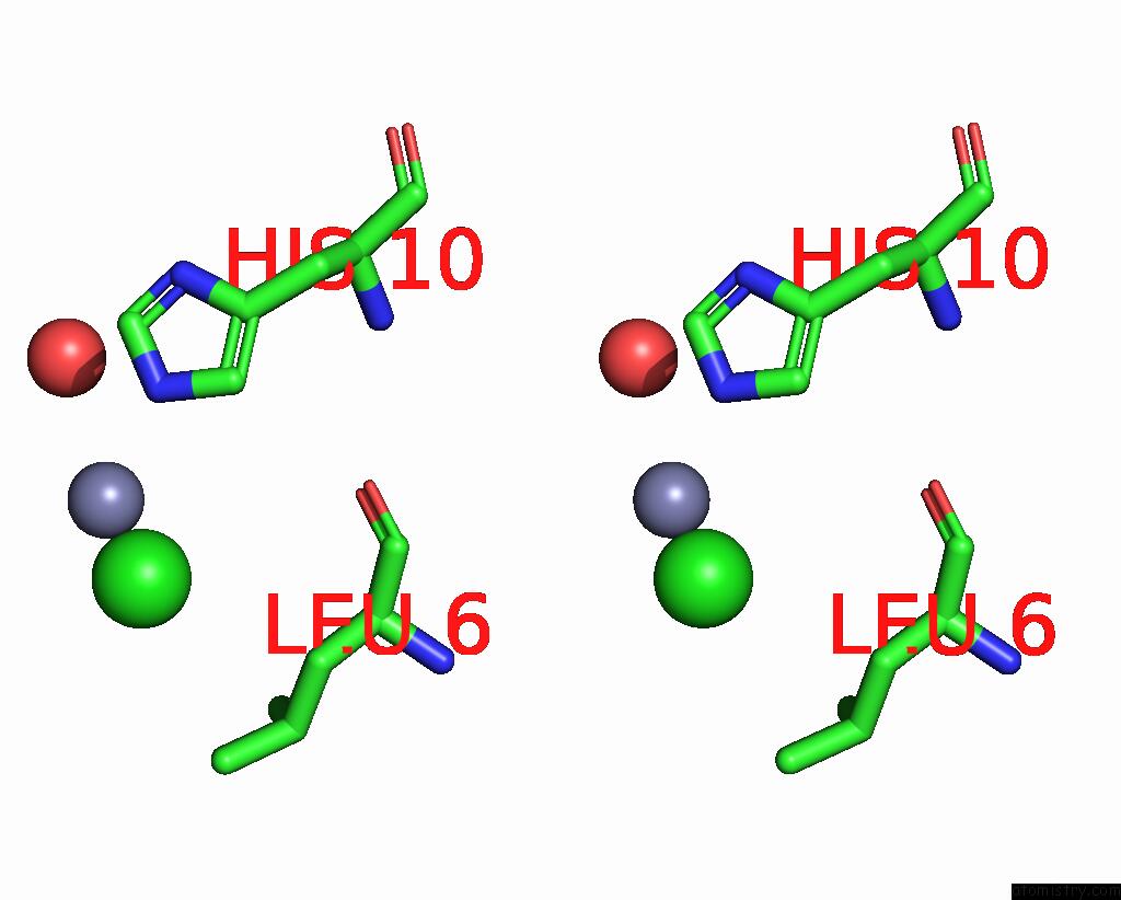
Zinc binding site 2 out of 4 in 8hgz
Go back to
Zinc binding site 2 out
of 4 in the Crystal Structure of Insulin
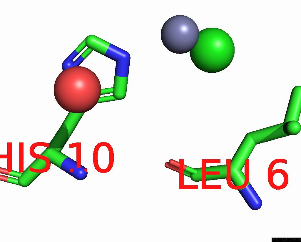
Mono view
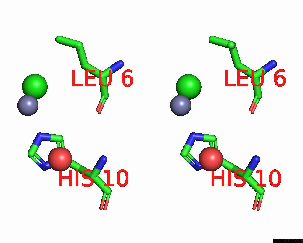
Stereo pair view
Mono view
Stereo pair view
A full contact list of Zinc with other atoms in the Zn binding
site number 2 of Crystal Structure of Insulin within 5.0Å range:
|
Zinc binding site 3 out of 4 in 8hgz
Go back to
Zinc binding site 3 out
of 4 in the Crystal Structure of Insulin
Mono view
Stereo pair view
Mono view
Stereo pair view
A full contact list of Zinc with other atoms in the Zn binding
site number 3 of Crystal Structure of Insulin within 5.0Å range:
|
Zinc binding site 4 out of 4 in 8hgz
Go back to
Zinc binding site 4 out
of 4 in the Crystal Structure of Insulin
Mono view
Stereo pair view
Mono view
Stereo pair view
A full contact list of Zinc with other atoms in the Zn binding
site number 4 of Crystal Structure of Insulin within 5.0Å range:
|
Reference:
E.Ayan,
E.Destan,
A.Kepceoglu,
H.I.Ciftci,
A.Kati,
H.Demirci.
Comparative Study of High-Resolution LYSB29(N Epsilon-Myristoyl) Des(B30) Insulin Structures Display Novel Dynamic Causal Interrelations in Monomeric-Dimeric Motions Crystals V. 13 2023.
ISSN: ESSN 2073-4352
DOI: 10.3390/CRYST13040648
Page generated: Thu Oct 31 07:16:17 2024
ISSN: ESSN 2073-4352
DOI: 10.3390/CRYST13040648
Last articles
Zn in 9MJ5Zn in 9HNW
Zn in 9G0L
Zn in 9FNE
Zn in 9DZN
Zn in 9E0I
Zn in 9D32
Zn in 9DAK
Zn in 8ZXC
Zn in 8ZUF