Zinc in PDB 8h5y: Crystal Structure of Radd- Adp Complex
Enzymatic activity of Crystal Structure of Radd- Adp Complex
All present enzymatic activity of Crystal Structure of Radd- Adp Complex:
3.6.4.12;
3.6.4.12;
Protein crystallography data
The structure of Crystal Structure of Radd- Adp Complex, PDB code: 8h5y
was solved by
X.X.Yan,
L.F.Tian,
with X-Ray Crystallography technique. A brief refinement statistics is given in the table below:
| Resolution Low / High (Å) | 19.96 / 2.70 |
| Space group | P 1 21 1 |
| Cell size a, b, c (Å), α, β, γ (°) | 82.089, 78.77, 110.238, 90, 99.57, 90 |
| R / Rfree (%) | 22.3 / 26.9 |
Other elements in 8h5y:
The structure of Crystal Structure of Radd- Adp Complex also contains other interesting chemical elements:
| Magnesium | (Mg) | 2 atoms |
Zinc Binding Sites:
The binding sites of Zinc atom in the Crystal Structure of Radd- Adp Complex
(pdb code 8h5y). This binding sites where shown within
5.0 Angstroms radius around Zinc atom.
In total 2 binding sites of Zinc where determined in the Crystal Structure of Radd- Adp Complex, PDB code: 8h5y:
Jump to Zinc binding site number: 1; 2;
In total 2 binding sites of Zinc where determined in the Crystal Structure of Radd- Adp Complex, PDB code: 8h5y:
Jump to Zinc binding site number: 1; 2;
Zinc binding site 1 out of 2 in 8h5y
Go back to
Zinc binding site 1 out
of 2 in the Crystal Structure of Radd- Adp Complex

Mono view
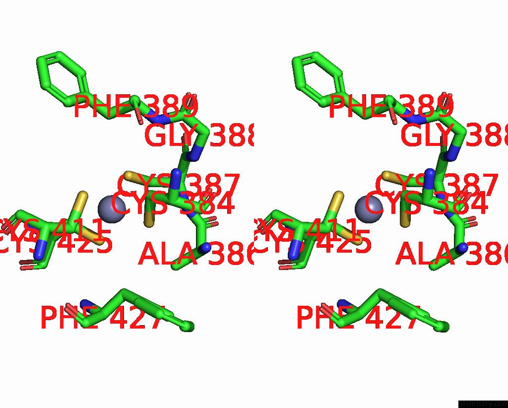
Stereo pair view
Mono view
Stereo pair view
A full contact list of Zinc with other atoms in the Zn binding
site number 1 of Crystal Structure of Radd- Adp Complex within 5.0Å range:
|
Zinc binding site 2 out of 2 in 8h5y
Go back to
Zinc binding site 2 out
of 2 in the Crystal Structure of Radd- Adp Complex

Mono view

Stereo pair view
Mono view
Stereo pair view
A full contact list of Zinc with other atoms in the Zn binding
site number 2 of Crystal Structure of Radd- Adp Complex within 5.0Å range:
|
Reference:
L.F.Tian,
X.Kuang,
K.Ding,
H.Gao,
Q.Tang,
X.X.Yan,
W.Xu.
Biochemical and Structural Analyses Shed Light on the Mechanisms of Radd Dna Binding and Its Atpase From Escherichia Coli. Int J Mol Sci V. 24 2023.
ISSN: ESSN 1422-0067
PubMed: 36614183
DOI: 10.3390/IJMS24010741
Page generated: Thu Oct 31 07:09:24 2024
ISSN: ESSN 1422-0067
PubMed: 36614183
DOI: 10.3390/IJMS24010741
Last articles
Zn in 9MJ5Zn in 9HNW
Zn in 9G0L
Zn in 9FNE
Zn in 9DZN
Zn in 9E0I
Zn in 9D32
Zn in 9DAK
Zn in 8ZXC
Zn in 8ZUF