Zinc in PDB 8e7b: Crystal Structure of the P53 (Y107H) Core Domain Monoclinic P Form
Protein crystallography data
The structure of Crystal Structure of the P53 (Y107H) Core Domain Monoclinic P Form, PDB code: 8e7b
was solved by
S.Lovell,
L.Liu,
K.P.Battaile,
S.Miller,
J.Karanicolas,
with X-Ray Crystallography technique. A brief refinement statistics is given in the table below:
| Resolution Low / High (Å) | 43.13 / 2.50 |
| Space group | P 1 21 1 |
| Cell size a, b, c (Å), α, β, γ (°) | 43.394, 68.636, 66.951, 90, 96.32, 90 |
| R / Rfree (%) | 21.9 / 27.9 |
Zinc Binding Sites:
The binding sites of Zinc atom in the Crystal Structure of the P53 (Y107H) Core Domain Monoclinic P Form
(pdb code 8e7b). This binding sites where shown within
5.0 Angstroms radius around Zinc atom.
In total 2 binding sites of Zinc where determined in the Crystal Structure of the P53 (Y107H) Core Domain Monoclinic P Form, PDB code: 8e7b:
Jump to Zinc binding site number: 1; 2;
In total 2 binding sites of Zinc where determined in the Crystal Structure of the P53 (Y107H) Core Domain Monoclinic P Form, PDB code: 8e7b:
Jump to Zinc binding site number: 1; 2;
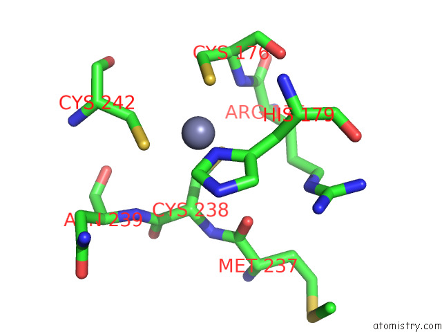
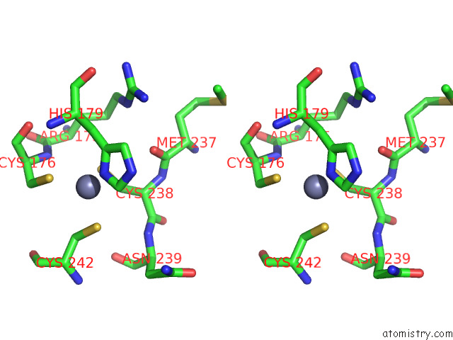
Zinc binding site 1 out of 2 in 8e7b
Go back to
Zinc binding site 1 out
of 2 in the Crystal Structure of the P53 (Y107H) Core Domain Monoclinic P Form
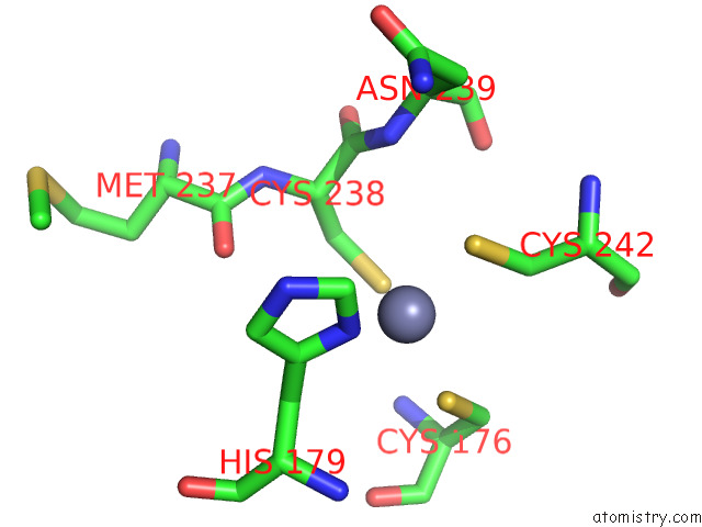
Mono view

Stereo pair view
Mono view
Stereo pair view
A full contact list of Zinc with other atoms in the Zn binding
site number 1 of Crystal Structure of the P53 (Y107H) Core Domain Monoclinic P Form within 5.0Å range:
|
Zinc binding site 2 out of 2 in 8e7b
Go back to
Zinc binding site 2 out
of 2 in the Crystal Structure of the P53 (Y107H) Core Domain Monoclinic P Form
Mono view
Stereo pair view
Mono view
Stereo pair view
A full contact list of Zinc with other atoms in the Zn binding
site number 2 of Crystal Structure of the P53 (Y107H) Core Domain Monoclinic P Form within 5.0Å range:
|
Reference:
A.Indeglia,
J.C.Leung,
S.A.Miller,
J.I.Leu,
J.F.Dougherty,
N.L.Clarke,
N.A.Kirven,
C.Shao,
L.Ke,
S.Lovell,
T.Barnoud,
D.Y.Lu,
C.Lin,
T.Kannan,
K.P.Battaile,
T.H.L.Yang,
I.Batista Oliva,
D.T.Claiborne,
P.Vogel,
L.Liu,
Q.Liu,
Y.Nefedova,
J.Cassel,
N.Auslander,
A.V.Kossenkov,
J.Karanicolas,
M.E.Murphy.
An African-Specific Variant of TP53 Reveals PADI4 As A Regulator of P53-Mediated Tumor Suppression. Cancer Discov 2023.
ISSN: ESSN 2159-8290
PubMed: 37140445
DOI: 10.1158/2159-8290.CD-22-1315
Page generated: Wed Oct 30 19:33:06 2024
ISSN: ESSN 2159-8290
PubMed: 37140445
DOI: 10.1158/2159-8290.CD-22-1315
Last articles
Zn in 9MJ5Zn in 9HNW
Zn in 9G0L
Zn in 9FNE
Zn in 9DZN
Zn in 9E0I
Zn in 9D32
Zn in 9DAK
Zn in 8ZXC
Zn in 8ZUF