Zinc in PDB 8aas: Crystal Structure of the Pyrococcus Abyssi Rpa Trimerization Core Bound to Poly-DT20 Ssdna
Protein crystallography data
The structure of Crystal Structure of the Pyrococcus Abyssi Rpa Trimerization Core Bound to Poly-DT20 Ssdna, PDB code: 8aas
was solved by
C.Madru,
P.Legrand,
L.Sauguet,
with X-Ray Crystallography technique. A brief refinement statistics is given in the table below:
| Resolution Low / High (Å) | 48.26 / 3.20 |
| Space group | P 32 2 1 |
| Cell size a, b, c (Å), α, β, γ (°) | 75.234, 75.234, 215.487, 90, 90, 120 |
| R / Rfree (%) | 26.4 / 29.3 |
Other elements in 8aas:
The structure of Crystal Structure of the Pyrococcus Abyssi Rpa Trimerization Core Bound to Poly-DT20 Ssdna also contains other interesting chemical elements:
| Calcium | (Ca) | 5 atoms |
Zinc Binding Sites:
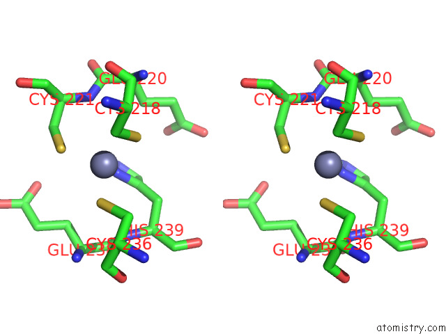
The binding sites of Zinc atom in the Crystal Structure of the Pyrococcus Abyssi Rpa Trimerization Core Bound to Poly-DT20 Ssdna
(pdb code 8aas). This binding sites where shown within
5.0 Angstroms radius around Zinc atom.
In total only one binding site of Zinc was determined in the Crystal Structure of the Pyrococcus Abyssi Rpa Trimerization Core Bound to Poly-DT20 Ssdna, PDB code: 8aas:
In total only one binding site of Zinc was determined in the Crystal Structure of the Pyrococcus Abyssi Rpa Trimerization Core Bound to Poly-DT20 Ssdna, PDB code: 8aas:
Zinc binding site 1 out of 1 in 8aas
Go back to
Zinc binding site 1 out
of 1 in the Crystal Structure of the Pyrococcus Abyssi Rpa Trimerization Core Bound to Poly-DT20 Ssdna

Mono view
Stereo pair view
Mono view
Stereo pair view
A full contact list of Zinc with other atoms in the Zn binding
site number 1 of Crystal Structure of the Pyrococcus Abyssi Rpa Trimerization Core Bound to Poly-DT20 Ssdna within 5.0Å range:
|
Reference:
C.Madru,
M.Martinez-Carranza,
S.Laurent,
A.C.Alberti,
M.Chevreuil,
B.Raynal,
A.Haouz,
R.A.Le Meur,
M.Delarue,
G.Henneke,
D.Flament,
M.Krupovic,
P.Legrand,
L.Sauguet.
Dna-Binding Mechanism and Evolution of Replication Protein A. Nat Commun V. 14 2326 2023.
ISSN: ESSN 2041-1723
PubMed: 37087464
DOI: 10.1038/S41467-023-38048-W
Page generated: Wed Oct 30 17:37:43 2024
ISSN: ESSN 2041-1723
PubMed: 37087464
DOI: 10.1038/S41467-023-38048-W
Last articles
Zn in 9MJ5Zn in 9HNW
Zn in 9G0L
Zn in 9FNE
Zn in 9DZN
Zn in 9E0I
Zn in 9D32
Zn in 9DAK
Zn in 8ZXC
Zn in 8ZUF