Zinc in PDB 7y3i: Structure of Dna Bound SALL4

Protein crystallography data

The structure of Structure of Dna Bound SALL4, PDB code: 7y3i was solved by W.Ru, C.Xu, with X-Ray Crystallography technique. A brief refinement statistics is given in the table below:

Resolution Low / High (Å) 43.13 / 2.45
Space group P 21 21 21
Cell size a, b, c (Å), α, β, γ (°) 48.975, 69.197, 110.295, 90, 90, 90
R / Rfree (%) 19.4 / 24.2

Zinc Binding Sites:

The binding sites of Zinc atom in the Structure of Dna Bound SALL4 (pdb code 7y3i). This binding sites where shown within 5.0 Angstroms radius around Zinc atom.
In total 6 binding sites of Zinc where determined in the Structure of Dna Bound SALL4, PDB code: 7y3i:
Jump to Zinc binding site number: 1; 2; 3; 4; 5; 6;

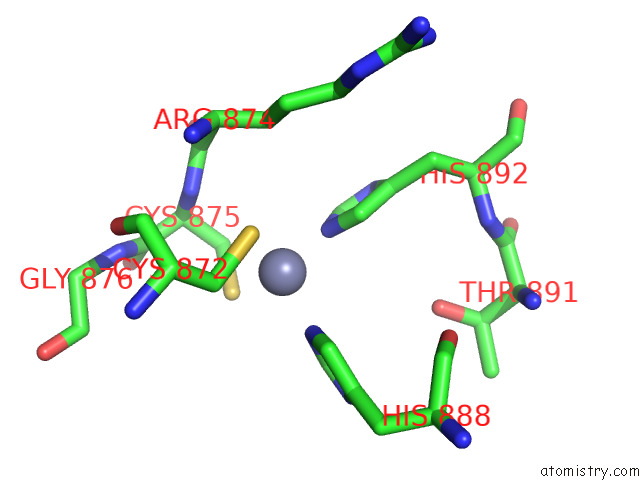
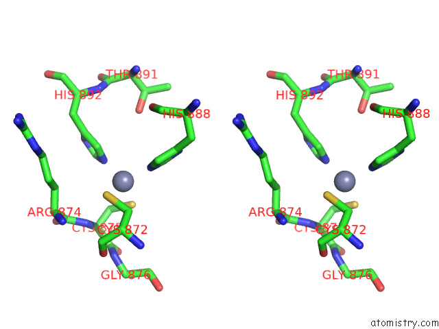
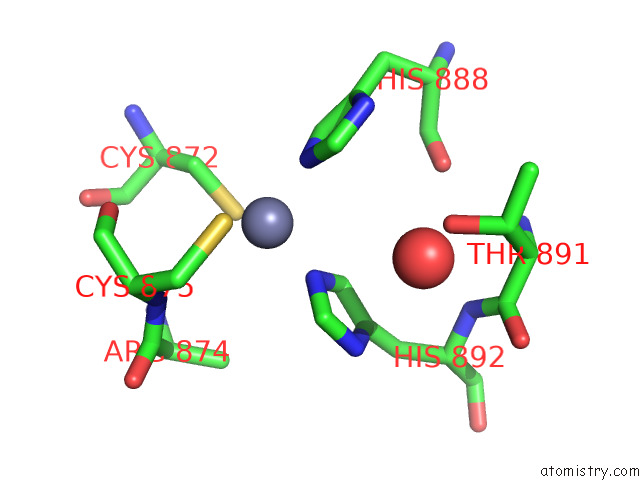
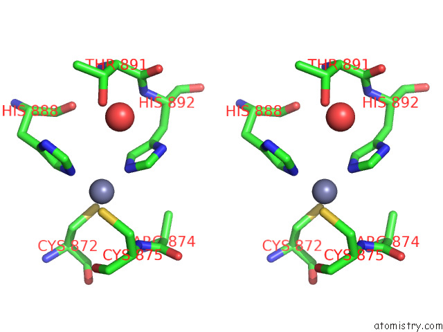
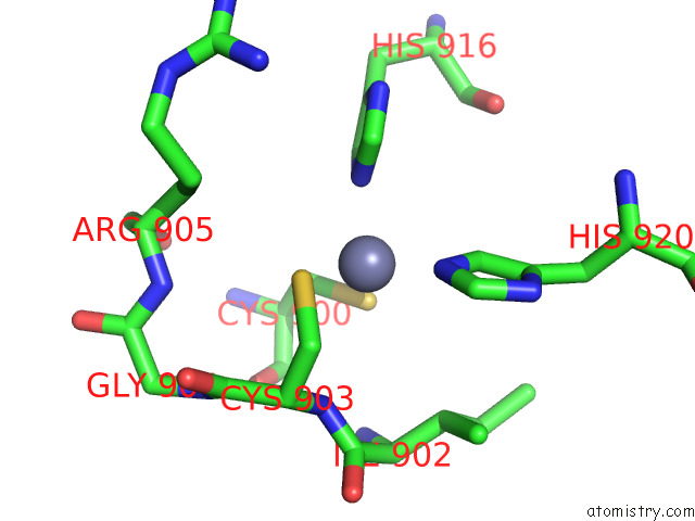
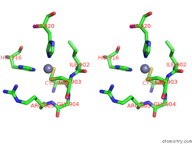
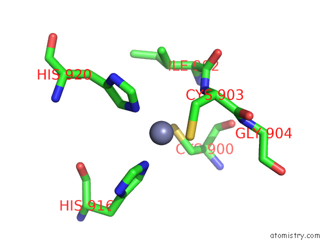
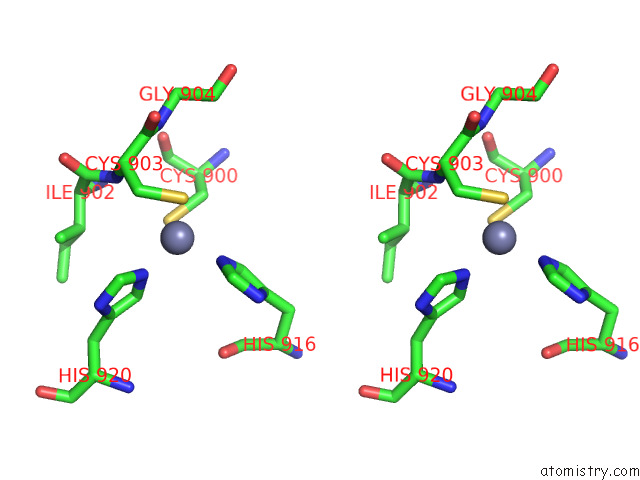
Zinc binding site 1 out of 6 in 7y3i

Go back to Zinc Binding Sites List in 7y3i
Zinc binding site 1 out of 6 in the Structure of Dna Bound SALL4


Mono view


Stereo pair view

A full contact list of Zinc with other atoms in the Zn binding site number 1 of Structure of Dna Bound SALL4 within 5.0Å range:
probe atom residue distance (Å) B Occ
A:Zn1001

b:30.4
occ:1.00
NE2 A:HIS892 1.9 45.5 1.0
NE2 A:HIS888 2.0 39.0 1.0
SG A:CYS875 2.2 37.6 1.0
SG A:CYS872 2.3 40.2 1.0
CD2 A:HIS892 2.8 43.6 1.0
CE1 A:HIS888 3.0 35.1 1.0
CE1 A:HIS892 3.0 43.4 1.0
CD2 A:HIS888 3.0 34.8 1.0
CB A:CYS872 3.2 37.8 1.0
CB A:CYS875 3.2 33.2 1.0
N A:CYS875 3.7 32.4 1.0
CG A:HIS892 4.0 42.7 1.0
CA A:CYS875 4.0 31.7 1.0
ND1 A:HIS892 4.1 41.8 1.0
ND1 A:HIS888 4.1 34.9 1.0
CG A:HIS888 4.2 32.8 1.0
CB A:ARG874 4.4 42.3 1.0
C A:ARG874 4.5 37.3 1.0
CA A:CYS872 4.6 37.2 1.0
C A:CYS875 4.7 32.5 1.0
CA A:ARG874 4.8 39.4 1.0
N A:ARG874 4.8 37.8 1.0
N A:GLY876 4.9 33.0 1.0
OG1 A:THR891 4.9 37.9 1.0

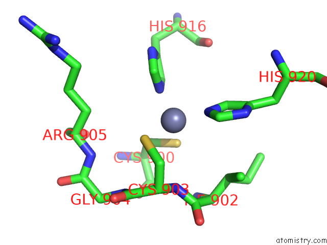
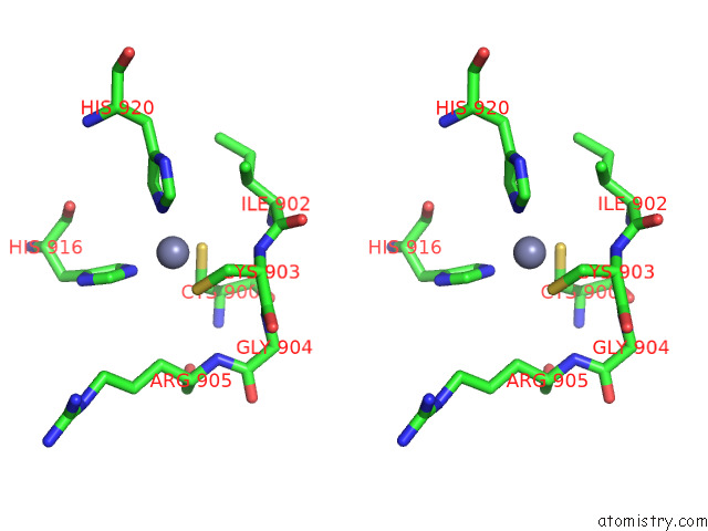
Zinc binding site 2 out of 6 in 7y3i

Go back to Zinc Binding Sites List in 7y3i
Zinc binding site 2 out of 6 in the Structure of Dna Bound SALL4


Mono view


Stereo pair view

A full contact list of Zinc with other atoms in the Zn binding site number 2 of Structure of Dna Bound SALL4 within 5.0Å range:
probe atom residue distance (Å) B Occ
A:Zn1002

b:32.7
occ:1.00
NE2 A:HIS916 2.1 40.5 1.0
NE2 A:HIS920 2.1 40.1 1.0
SG A:CYS900 2.3 33.0 1.0
SG A:CYS903 2.3 40.0 1.0
CD2 A:HIS916 2.9 38.8 1.0
CE1 A:HIS920 3.0 36.1 1.0
CD2 A:HIS920 3.1 38.3 1.0
CB A:CYS900 3.1 32.6 1.0
CE1 A:HIS916 3.1 38.7 1.0
CB A:CYS903 3.1 37.2 1.0
N A:CYS903 3.6 33.0 1.0
CA A:CYS903 3.9 34.8 1.0
CG A:HIS916 4.1 37.4 1.0
ND1 A:HIS920 4.1 38.8 1.0
ND1 A:HIS916 4.1 37.4 1.0
CG A:HIS920 4.2 39.9 1.0
CB A:ILE902 4.5 40.3 1.0
C A:CYS903 4.5 34.7 1.0
CA A:CYS900 4.6 31.6 1.0
C A:ILE902 4.6 31.9 1.0
N A:GLY904 4.7 32.2 1.0
CB A:ARG905 4.8 45.1 1.0
CG2 A:ILE902 4.9 26.6 1.0
N A:ARG905 4.9 35.6 1.0
CA A:ILE902 4.9 34.1 1.0
N A:ILE902 5.0 31.5 1.0

Zinc binding site 3 out of 6 in 7y3i

Go back to Zinc Binding Sites List in 7y3i
Zinc binding site 3 out of 6 in the Structure of Dna Bound SALL4


Mono view


Stereo pair view

A full contact list of Zinc with other atoms in the Zn binding site number 3 of Structure of Dna Bound SALL4 within 5.0Å range:
probe atom residue distance (Å) B Occ
B:Zn1001

b:36.1
occ:1.00
NE2 B:HIS892 2.0 46.0 1.0
NE2 B:HIS888 2.0 45.5 1.0
SG B:CYS875 2.3 37.4 1.0
SG B:CYS872 2.4 44.5 1.0
CE1 B:HIS892 3.0 42.0 1.0
CD2 B:HIS892 3.0 42.6 1.0
CE1 B:HIS888 3.0 40.8 1.0
CD2 B:HIS888 3.0 41.6 1.0
CB B:CYS872 3.1 42.6 1.0
N B:CYS875 3.5 46.4 1.0
CB B:CYS875 3.6 40.2 1.0
CA B:CYS875 4.0 47.0 1.0
ND1 B:HIS892 4.1 41.6 1.0
ND1 B:HIS888 4.1 39.7 1.0
CG B:HIS892 4.1 41.7 1.0
CG B:HIS888 4.2 39.6 1.0
O B:CYS875 4.2 59.6 1.0
C B:CYS875 4.4 55.0 1.0
C B:ARG874 4.5 47.6 1.0
CB B:ARG874 4.5 45.2 1.0
CA B:CYS872 4.6 49.4 1.0
CA B:ARG874 4.9 49.4 1.0
N B:ARG874 4.9 53.6 1.0
OG1 B:THR891 4.9 34.2 1.0
O B:HOH1103 5.0 36.3 1.0

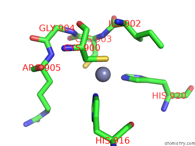
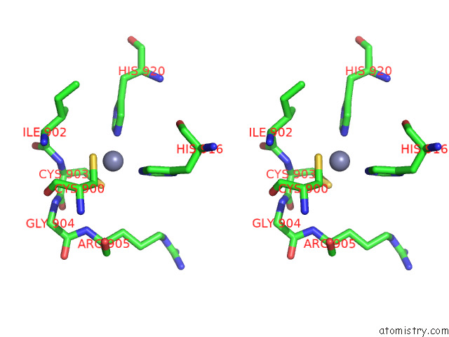
Zinc binding site 4 out of 6 in 7y3i

Go back to Zinc Binding Sites List in 7y3i
Zinc binding site 4 out of 6 in the Structure of Dna Bound SALL4


Mono view


Stereo pair view

A full contact list of Zinc with other atoms in the Zn binding site number 4 of Structure of Dna Bound SALL4 within 5.0Å range:
probe atom residue distance (Å) B Occ
B:Zn1002

b:26.7
occ:1.00
NE2 B:HIS916 2.0 31.7 1.0
NE2 B:HIS920 2.1 36.5 1.0
SG B:CYS900 2.3 24.0 1.0
SG B:CYS903 2.3 26.7 1.0
CE1 B:HIS916 2.9 28.3 1.0
CD2 B:HIS920 3.0 36.4 1.0
CD2 B:HIS916 3.1 28.0 1.0
CE1 B:HIS920 3.1 34.5 1.0
CB B:CYS900 3.2 24.8 1.0
CB B:CYS903 3.4 21.8 1.0
N B:CYS903 3.7 27.6 1.0
ND1 B:HIS916 4.0 29.5 1.0
CA B:CYS903 4.1 23.1 1.0
CG B:HIS916 4.1 28.5 1.0
ND1 B:HIS920 4.1 36.1 1.0
CG B:HIS920 4.2 37.5 1.0
CB B:ILE902 4.4 29.5 1.0
C B:ILE902 4.6 27.0 1.0
CA B:CYS900 4.6 25.8 1.0
C B:CYS903 4.7 26.0 1.0
CG2 B:ILE902 4.7 26.4 1.0
N B:GLY904 4.7 22.5 1.0
CB B:ARG905 4.7 23.7 1.0
N B:ARG905 4.9 23.2 1.0
CA B:ILE902 4.9 27.1 1.0
N B:ILE902 4.9 23.9 1.0

Zinc binding site 5 out of 6 in 7y3i

Go back to Zinc Binding Sites List in 7y3i
Zinc binding site 5 out of 6 in the Structure of Dna Bound SALL4


Mono view


Stereo pair view

A full contact list of Zinc with other atoms in the Zn binding site number 5 of Structure of Dna Bound SALL4 within 5.0Å range:
probe atom residue distance (Å) B Occ
C:Zn1001

b:29.1
occ:0.99
NE2 C:HIS916 2.1 36.2 1.0
SG C:CYS903 2.2 38.2 1.0
NE2 C:HIS920 2.2 34.6 1.0
SG C:CYS900 2.3 30.9 1.0
CD2 C:HIS920 2.7 31.2 1.0
CE1 C:HIS916 3.1 33.3 1.0
CB C:CYS900 3.1 29.6 1.0
CD2 C:HIS916 3.1 35.1 1.0
CB C:CYS903 3.3 33.7 1.0
CE1 C:HIS920 3.4 33.8 1.0
N C:CYS903 3.6 32.7 1.0
CG C:HIS920 4.0 32.6 1.0
CA C:CYS903 4.0 33.8 1.0
ND1 C:HIS916 4.2 34.4 1.0
CG C:HIS916 4.2 35.1 1.0
ND1 C:HIS920 4.3 33.8 1.0
CB C:ILE902 4.3 35.0 1.0
CA C:CYS900 4.5 28.2 1.0
C C:ILE902 4.5 34.0 1.0
C C:CYS903 4.7 35.1 1.0
CB C:ARG905 4.7 35.3 1.0
O C:CYS900 4.8 28.5 1.0
CA C:ILE902 4.8 33.6 1.0
N C:ILE902 4.8 31.6 1.0
C C:CYS900 4.8 29.2 1.0
N C:GLY904 4.9 33.4 1.0
CG2 C:ILE902 4.9 28.5 1.0
CG C:ARG905 4.9 36.4 1.0
N C:ARG905 5.0 29.6 1.0

Zinc binding site 6 out of 6 in 7y3i

Go back to Zinc Binding Sites List in 7y3i
Zinc binding site 6 out of 6 in the Structure of Dna Bound SALL4


Mono view


Stereo pair view

A full contact list of Zinc with other atoms in the Zn binding site number 6 of Structure of Dna Bound SALL4 within 5.0Å range:
probe atom residue distance (Å) B Occ
D:Zn1001

b:41.8
occ:1.00
NE2 D:HIS916 2.1 55.1 1.0
NE2 D:HIS920 2.1 70.5 1.0
SG D:CYS900 2.3 34.2 1.0
SG D:CYS903 2.4 39.5 1.0
CD2 D:HIS920 2.9 71.0 1.0
CE1 D:HIS916 3.0 52.4 1.0
CD2 D:HIS916 3.1 52.0 1.0
CB D:CYS903 3.2 41.4 1.0
CB D:CYS900 3.2 30.3 1.0
CE1 D:HIS920 3.2 70.7 1.0
N D:CYS903 3.5 43.0 1.0
CA D:CYS903 3.8 42.4 1.0
ND1 D:HIS916 4.1 52.9 1.0
CG D:HIS920 4.1 71.3 1.0
CG D:HIS916 4.2 51.8 1.0
ND1 D:HIS920 4.3 71.5 1.0
CB D:ILE902 4.5 43.6 1.0
C D:CYS903 4.5 42.2 1.0
C D:ILE902 4.6 42.7 1.0
CA D:CYS900 4.6 32.3 1.0
N D:GLY904 4.7 37.1 1.0
CG2 D:ILE902 4.8 41.5 1.0
CA D:ILE902 4.9 42.1 1.0
N D:ILE902 5.0 39.5 1.0
C D:CYS900 5.0 37.7 1.0

Reference:

W.Ru, T.Koga, X.Wang, Q.Guo, M.D.Gearhart, S.Zhao, M.Murphy, H.Kawakami, D.Corcoran, J.Zhang, Z.Zhu, X.Yao, Y.Kawakami, C.Xu. Structural Studies of Sall Family Protein Zinc Finger Cluster Domains in Complex with Dna Reveal Preferential Binding to An Aata Tetranucleotide Motif. J.Biol.Chem. V. 298 02607 2022.
ISSN: ESSN 1083-351X
PubMed: 36257403
DOI: 10.1016/J.JBC.2022.102607
Page generated: Sat Apr 8 06:05:34 2023

Last articles

Zn in 8WB0
Zn in 8WAX
Zn in 8WAU
Zn in 8WAZ
Zn in 8WAY
Zn in 8WAV
Zn in 8WAW
Zn in 8WAT
Zn in 8W7M
Zn in 8WD3
© Copyright 2008-2020 by atomistry.com
Home   |    Site Map   |    Copyright   |    Contact us   |    Privacy