Zinc in PDB 7xp0: Crystal Structure of Pmir From Pseudomonas Aeruginosa
Protein crystallography data
The structure of Crystal Structure of Pmir From Pseudomonas Aeruginosa, PDB code: 7xp0
was solved by
Y.X.Zhang,
H.H.Liang,
J.H.Gan,
with X-Ray Crystallography technique. A brief refinement statistics is given in the table below:
| Resolution Low / High (Å) | 29.38 / 2.61 |
| Space group | I 41 2 2 |
| Cell size a, b, c (Å), α, β, γ (°) | 74.352, 74.352, 188.238, 90, 90, 90 |
| R / Rfree (%) | 20 / 25 |
Zinc Binding Sites:
The binding sites of Zinc atom in the Crystal Structure of Pmir From Pseudomonas Aeruginosa
(pdb code 7xp0). This binding sites where shown within
5.0 Angstroms radius around Zinc atom.
In total only one binding site of Zinc was determined in the Crystal Structure of Pmir From Pseudomonas Aeruginosa, PDB code: 7xp0:
In total only one binding site of Zinc was determined in the Crystal Structure of Pmir From Pseudomonas Aeruginosa, PDB code: 7xp0:
Zinc binding site 1 out of 1 in 7xp0
Go back to
Zinc binding site 1 out
of 1 in the Crystal Structure of Pmir From Pseudomonas Aeruginosa
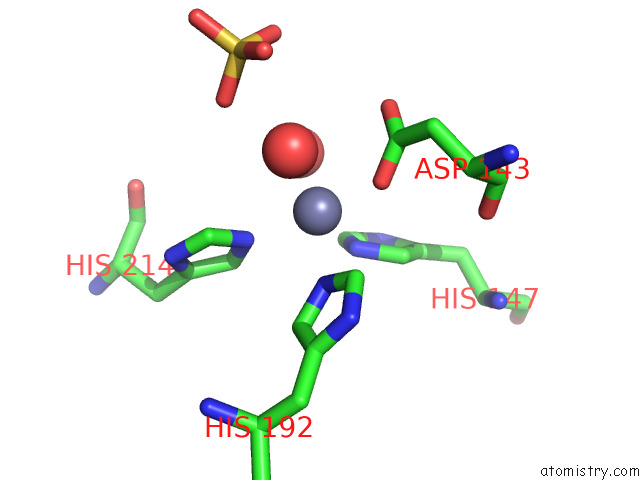
Mono view
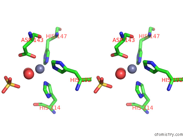
Stereo pair view
Mono view
Stereo pair view
A full contact list of Zinc with other atoms in the Zn binding
site number 1 of Crystal Structure of Pmir From Pseudomonas Aeruginosa within 5.0Å range:
|
Reference:
G.Cui,
Y.Zhang,
X.Xu,
Y.Liu,
Z.Li,
M.Wu,
J.Liu,
J.Gan,
H.Liang.
Pmir Senses 2-Methylisocitrate Levels to Regulate Bacterial Virulence in Pseudomonas Aeruginosa. Sci Adv V. 8 D4220 2022.
ISSN: ESSN 2375-2548
PubMed: 36475801
DOI: 10.1126/SCIADV.ADD4220
Page generated: Wed Oct 30 15:10:54 2024
ISSN: ESSN 2375-2548
PubMed: 36475801
DOI: 10.1126/SCIADV.ADD4220
Last articles
Zn in 9MJ5Zn in 9HNW
Zn in 9G0L
Zn in 9FNE
Zn in 9DZN
Zn in 9E0I
Zn in 9D32
Zn in 9DAK
Zn in 8ZXC
Zn in 8ZUF