Zinc in PDB 7x97: Crystal Structure of Actinomycin D-Echinomycin-D(Agcccgt/Acgggct) Complex
Protein crystallography data
The structure of Crystal Structure of Actinomycin D-Echinomycin-D(Agcccgt/Acgggct) Complex, PDB code: 7x97
was solved by
S.H.Kao,
R.B.Satange,
M.H.Hou,
with X-Ray Crystallography technique. A brief refinement statistics is given in the table below:
| Resolution Low / High (Å) | 25.57 / 1.95 |
| Space group | P 63 2 2 |
| Cell size a, b, c (Å), α, β, γ (°) | 43.452, 43.452, 139.365, 90, 90, 120 |
| R / Rfree (%) | 22.9 / 28.2 |
Other elements in 7x97:
The structure of Crystal Structure of Actinomycin D-Echinomycin-D(Agcccgt/Acgggct) Complex also contains other interesting chemical elements:
| Potassium | (K) | 1 atom |
| Chlorine | (Cl) | 2 atoms |
Zinc Binding Sites:
The binding sites of Zinc atom in the Crystal Structure of Actinomycin D-Echinomycin-D(Agcccgt/Acgggct) Complex
(pdb code 7x97). This binding sites where shown within
5.0 Angstroms radius around Zinc atom.
In total 5 binding sites of Zinc where determined in the Crystal Structure of Actinomycin D-Echinomycin-D(Agcccgt/Acgggct) Complex, PDB code: 7x97:
Jump to Zinc binding site number: 1; 2; 3; 4; 5;
In total 5 binding sites of Zinc where determined in the Crystal Structure of Actinomycin D-Echinomycin-D(Agcccgt/Acgggct) Complex, PDB code: 7x97:
Jump to Zinc binding site number: 1; 2; 3; 4; 5;
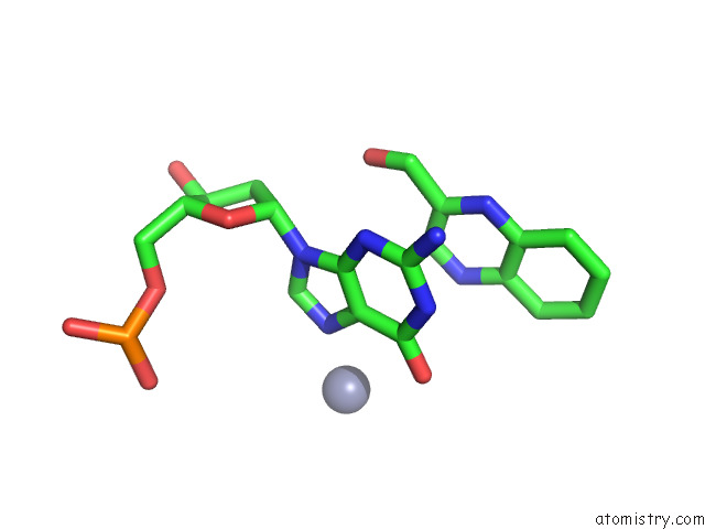
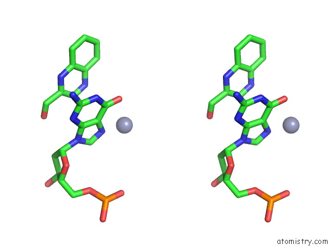
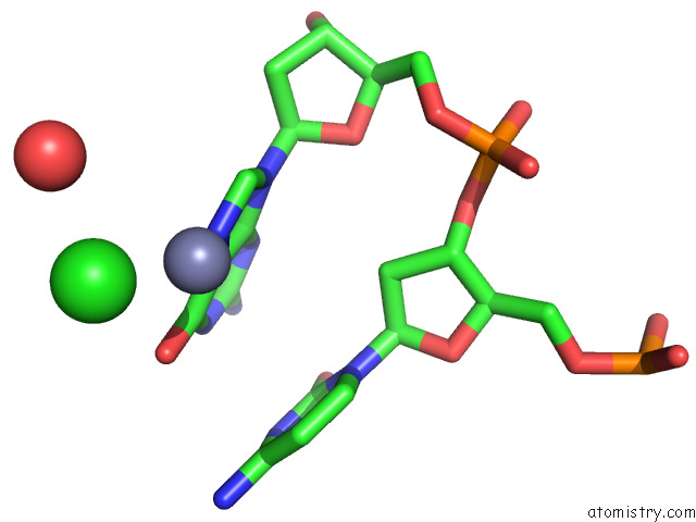
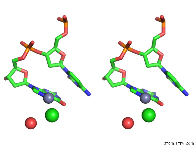
Zinc binding site 1 out of 5 in 7x97
Go back to
Zinc binding site 1 out
of 5 in the Crystal Structure of Actinomycin D-Echinomycin-D(Agcccgt/Acgggct) Complex
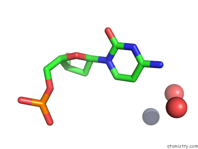
Mono view
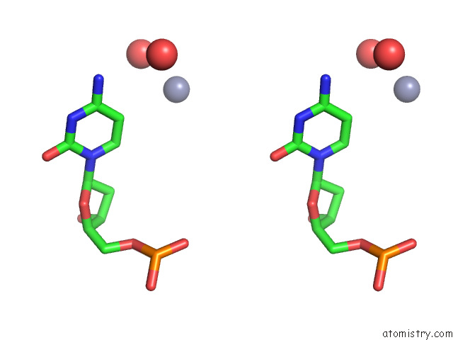
Stereo pair view
Mono view
Stereo pair view
A full contact list of Zinc with other atoms in the Zn binding
site number 1 of Crystal Structure of Actinomycin D-Echinomycin-D(Agcccgt/Acgggct) Complex within 5.0Å range:
|
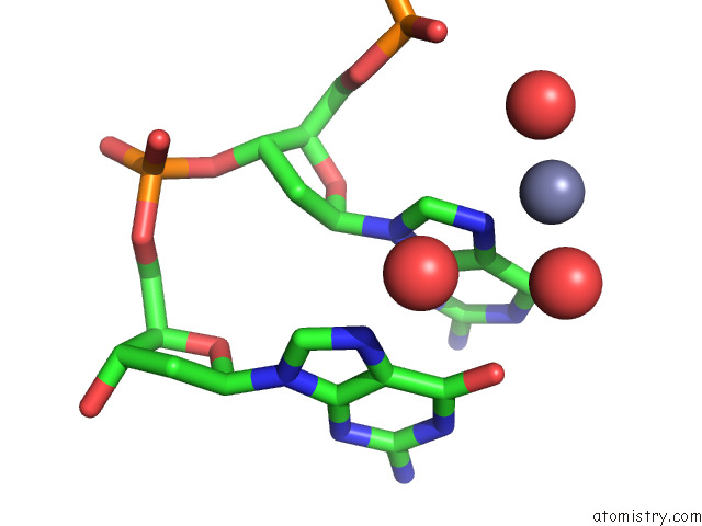
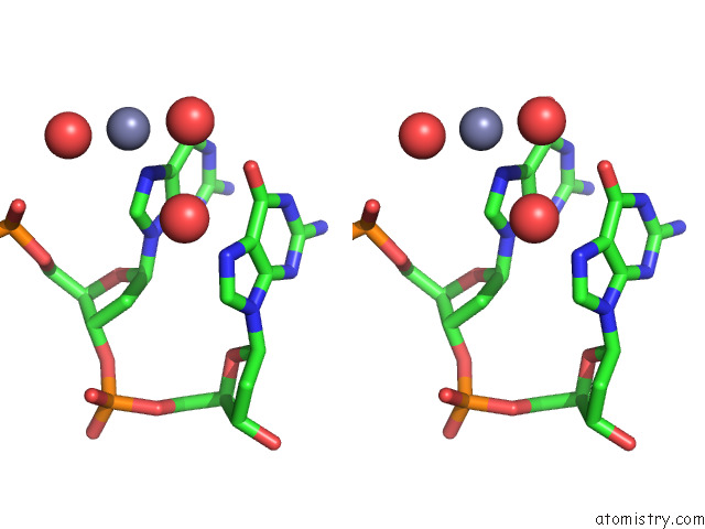
Zinc binding site 2 out of 5 in 7x97
Go back to
Zinc binding site 2 out
of 5 in the Crystal Structure of Actinomycin D-Echinomycin-D(Agcccgt/Acgggct) Complex
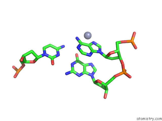
Mono view
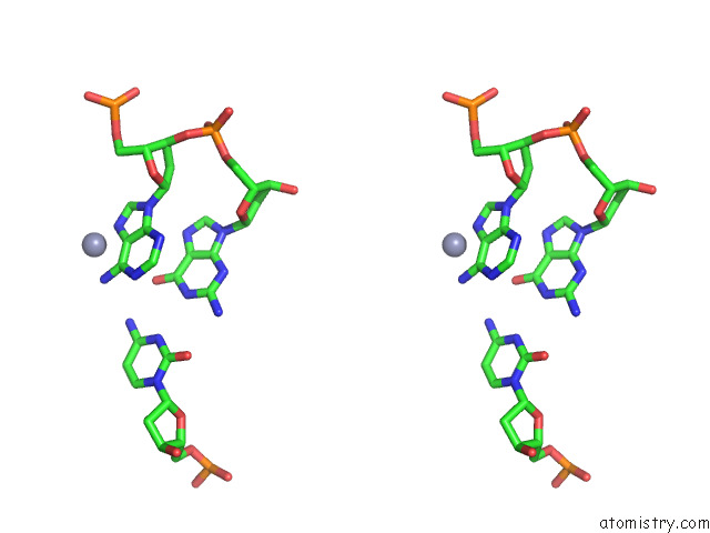
Stereo pair view
Mono view
Stereo pair view
A full contact list of Zinc with other atoms in the Zn binding
site number 2 of Crystal Structure of Actinomycin D-Echinomycin-D(Agcccgt/Acgggct) Complex within 5.0Å range:
|
Zinc binding site 3 out of 5 in 7x97
Go back to
Zinc binding site 3 out
of 5 in the Crystal Structure of Actinomycin D-Echinomycin-D(Agcccgt/Acgggct) Complex
Mono view
Stereo pair view
Mono view
Stereo pair view
A full contact list of Zinc with other atoms in the Zn binding
site number 3 of Crystal Structure of Actinomycin D-Echinomycin-D(Agcccgt/Acgggct) Complex within 5.0Å range:
|
Zinc binding site 4 out of 5 in 7x97
Go back to
Zinc binding site 4 out
of 5 in the Crystal Structure of Actinomycin D-Echinomycin-D(Agcccgt/Acgggct) Complex
Mono view
Stereo pair view
Mono view
Stereo pair view
A full contact list of Zinc with other atoms in the Zn binding
site number 4 of Crystal Structure of Actinomycin D-Echinomycin-D(Agcccgt/Acgggct) Complex within 5.0Å range:
|
Zinc binding site 5 out of 5 in 7x97
Go back to
Zinc binding site 5 out
of 5 in the Crystal Structure of Actinomycin D-Echinomycin-D(Agcccgt/Acgggct) Complex
Mono view
Stereo pair view
Mono view
Stereo pair view
A full contact list of Zinc with other atoms in the Zn binding
site number 5 of Crystal Structure of Actinomycin D-Echinomycin-D(Agcccgt/Acgggct) Complex within 5.0Å range:
|
Reference:
R.Satange,
S.H.Kao,
C.M.Chien,
S.H.Chou,
C.C.Lin,
S.Neidle,
M.H.Hou.
Staggered Intercalation of Dna Duplexes with Base-Pair Modulation By Two Distinct Drug Molecules Induces Asymmetric Backbone Twisting and Structure Polymorphism. Nucleic Acids Res. V. 50 8867 2022.
ISSN: ESSN 1362-4962
PubMed: 35871296
DOI: 10.1093/NAR/GKAC629
Page generated: Wed Oct 30 14:51:53 2024
ISSN: ESSN 1362-4962
PubMed: 35871296
DOI: 10.1093/NAR/GKAC629
Last articles
Zn in 9MJ5Zn in 9HNW
Zn in 9G0L
Zn in 9FNE
Zn in 9DZN
Zn in 9E0I
Zn in 9D32
Zn in 9DAK
Zn in 8ZXC
Zn in 8ZUF