Zinc in PDB 7wg4: Dvaa-KLATE1
Enzymatic activity of Dvaa-KLATE1
All present enzymatic activity of Dvaa-KLATE1:
2.3.2.8;
2.3.2.8;
Protein crystallography data
The structure of Dvaa-KLATE1, PDB code: 7wg4
was solved by
M.K.Kim,
B.H.Kim,
S.-J.Oh,
H.K.Song,
with X-Ray Crystallography technique. A brief refinement statistics is given in the table below:
| Resolution Low / High (Å) | 28.57 / 1.51 |
| Space group | P 43 21 2 |
| Cell size a, b, c (Å), α, β, γ (°) | 86.46, 86.46, 160.61, 90, 90, 90 |
| R / Rfree (%) | 18.5 / 21.6 |
Zinc Binding Sites:
The binding sites of Zinc atom in the Dvaa-KLATE1
(pdb code 7wg4). This binding sites where shown within
5.0 Angstroms radius around Zinc atom.
In total only one binding site of Zinc was determined in the Dvaa-KLATE1, PDB code: 7wg4:
In total only one binding site of Zinc was determined in the Dvaa-KLATE1, PDB code: 7wg4:
Zinc binding site 1 out of 1 in 7wg4
Go back to
Zinc binding site 1 out
of 1 in the Dvaa-KLATE1
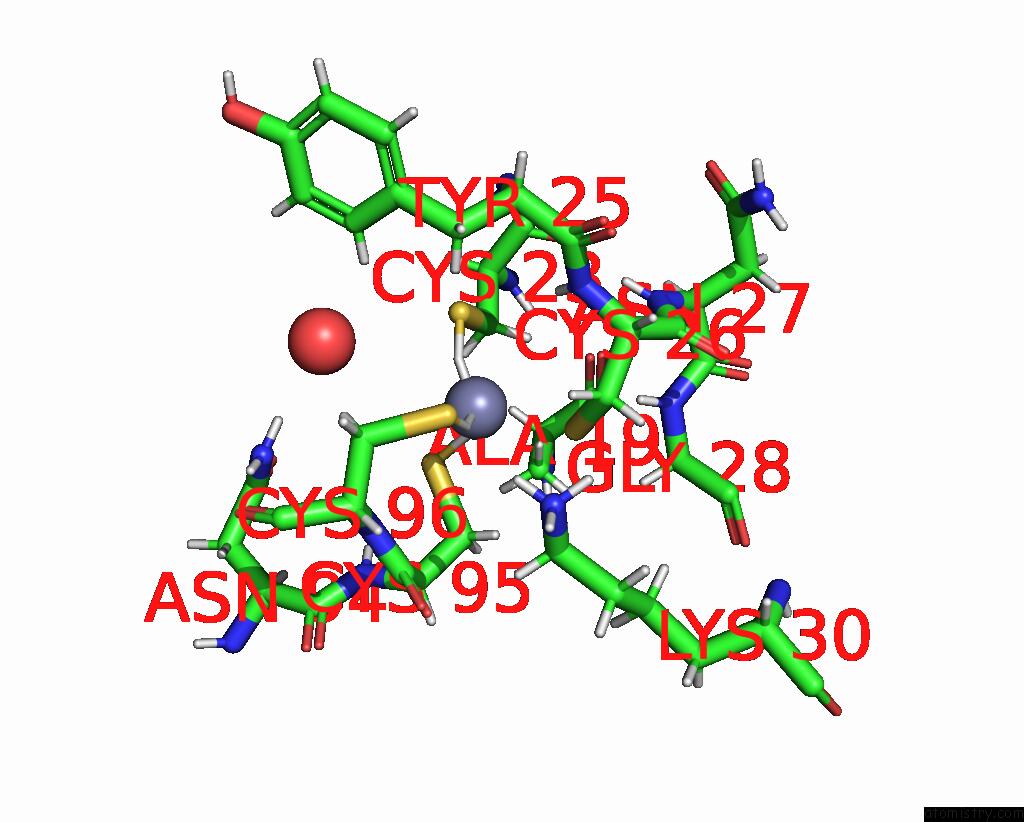
Mono view
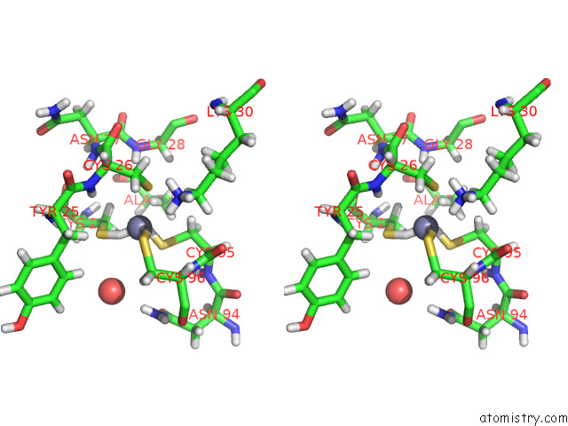
Stereo pair view
Mono view
Stereo pair view
A full contact list of Zinc with other atoms in the Zn binding
site number 1 of Dvaa-KLATE1 within 5.0Å range:
|
Reference:
B.H.Kim,
M.K.Kim,
S.J.Oh,
K.T.Nguyen,
J.H.Kim,
A.Varshavsky,
C.S.Hwang,
H.K.Song.
Crystal Structure of the ATE1 Arginyl-Trna-Protein Transferase and Arginylation of N-Degron Substrates. Proc.Natl.Acad.Sci.Usa V. 119 97119 2022.
ISSN: ESSN 1091-6490
PubMed: 35878037
DOI: 10.1073/PNAS.2209597119
Page generated: Wed Oct 30 14:24:29 2024
ISSN: ESSN 1091-6490
PubMed: 35878037
DOI: 10.1073/PNAS.2209597119
Last articles
Zn in 9MJ5Zn in 9HNW
Zn in 9G0L
Zn in 9FNE
Zn in 9DZN
Zn in 9E0I
Zn in 9D32
Zn in 9DAK
Zn in 8ZXC
Zn in 8ZUF